
MCA dot sign (brain)
Citation, DOI, disclosures and article data
At the time the article was created Frank Gaillard had no recorded disclosures.
View Frank Gaillard's current disclosuresAt the time the article was last revised Rohit Sharma had no financial relationships to ineligible companies to disclose.
View Rohit Sharma's current disclosures- Middle cerebral artery dot sign
- Middle cerebral artery (MCA) dot sign
- Middle cerebral arterial dot sign
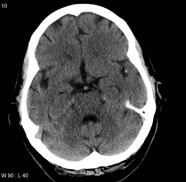
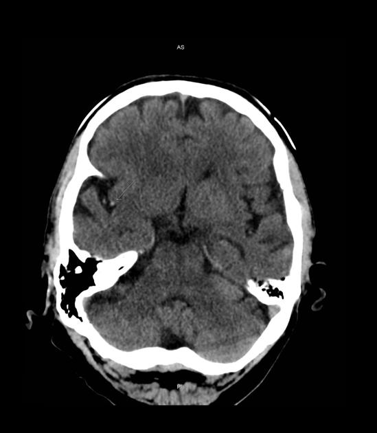
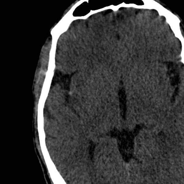
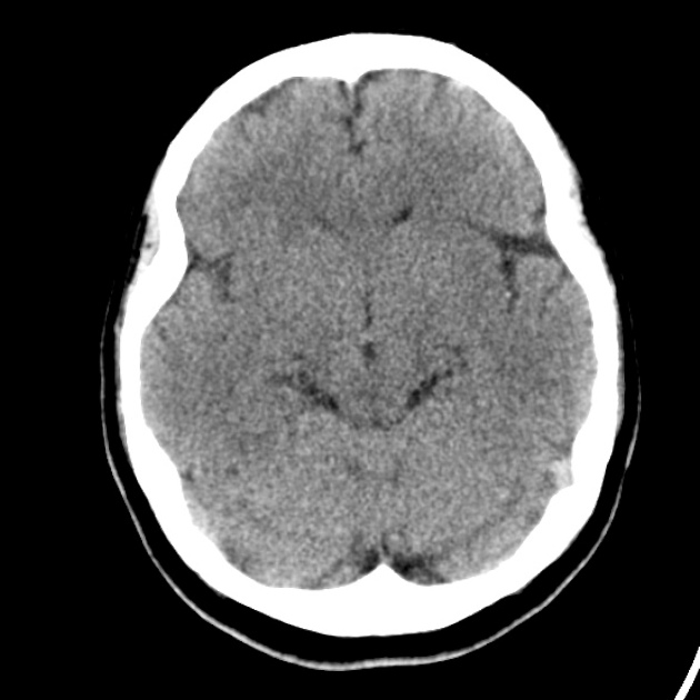
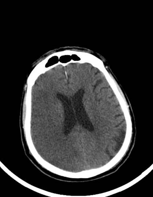
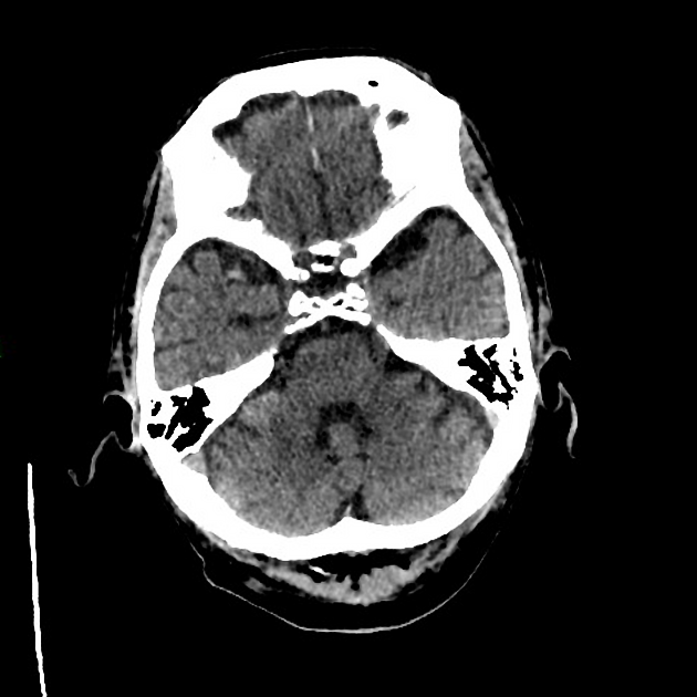
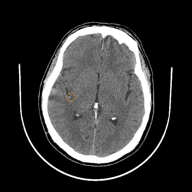
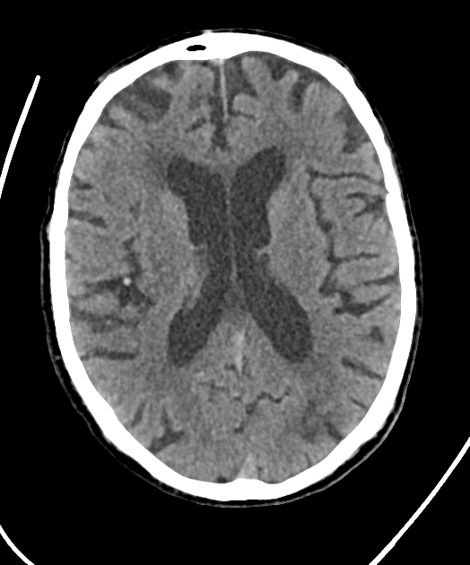
The middle cerebral artery (MCA) dot sign, also known as the Sylvian fissure sign, is a type of hyperdense vessel sign seen on non-contrast brain CT and represents the cross-sectional M2 equivalent of the hyperdense MCA sign. Rather than imaging a length of middle cerebral artery (typically the M1 segment), the dot sign represents a thromboembolism within a segmental branch of the MCA located within the Sylvian fissure (M2 segment). The sign appears when the high-attenuation MCA is viewed in axial section, since the occluded vessel courses in a plane perpendicular to the transverse plane of imaging.
Pathology
The MCA dot sign is an early marker of thromboembolic occlusion of the distal MCA branches seen in the Sylvian fissure (M2 segment). The principally affected area of the brain is the insula.
Sensitivity and specificity
The sensitivity of this CT sign is approximately 35%, while its specificity may be as high as 100% 3.
Treatment and prognosis
The MCA dot sign is prognostic of poor outcome in patients with ischaemic stroke, but with a better outcome than the hyperdense MCA sign 2.
As such, single studies have assessed the possibility of automated detection 4 and its value in prediction of secondary symptomatic intracranial haemorrhage 5.
Punctate vascular calcification along the M2 segment of the MCA within the Sylvian fissure represents a potential mimic of this sign.
References
- 1. Shetty SK. The MCA Dot Sign. Radiology. 2006;241 (1): 315-8. doi:10.1148/radiol.2411040573 - Pubmed citation
- 2. Barber PA, Demchuk AM, Hudon ME et-al. Hyperdense sylvian fissure MCA "dot" sign: A CT marker of acute ischemia. Stroke. 2001;32 (1): 84-8. Pubmed citation
- 3. Leary MC, Kidwell CS, Villablanca JP et-al. Validation of computed tomographic middle cerebral artery "dot"sign: an angiographic correlation study. Stroke. 2003;34 (11): 2636-40. doi:10.1161/01.STR.0000092123.00938.83 - Pubmed citation
- 4. Takahashi N, Lee Y, Tsai DY et-al. An automated detection method for the MCA dot sign of acute stroke in unenhanced CT. Radiol Phys Technol. 2014;7 (1): 79-88. doi:10.1007/s12194-013-0234-1 - Pubmed citation
- 5. Bentley P, Ganesalingam J, Carlton Jones AL et-al. Prediction of stroke thrombolysis outcome using CT brain machine learning. Neuroimage Clin. 2014;4: 635-40. doi:10.1016/j.nicl.2014.02.003 - Free text at pubmed - Pubmed citation
Incoming Links
- MCA territory acute infarction with hyperdense MCA sign
- Left MCA territory acute ischemic stroke with hyperdense MCA sign
- Dense left MCA sign with left MCA infarction
- Acute MCA stroke
- Acute brain infarction
- MCA dot sign
- MCA dot sign and haemorrhagic transformation of infarct
- Acute P1 occlusion with PCA ischaemic penumbra (CT perfusion)
- Acute A3 occlusion with ACA ischaemic penumbra (CT perfusion)
- Acute M1 occlusion with ischaemic penumbra (CT perfusion)
- MCA ischemic stroke - added value of CTA source image ASPECTS
- Haemorrhagic transformation of MCA infarct
- Acute middle cerebral artery territory infarct
- Middle cerebral artery territory infarct
- Middle cerebral artery dot sign
- Hyperdense MCA sign
- Hyperdense MCA sign
- Acute MCA infarction
Related articles: Stroke and intracranial haemorrhage
-
stroke and intracranial haemorrhage
- general articles
-
ischaemic stroke
- general discussions
- scoring and classification systems
- Alberta stroke program early CT score (ASPECTS)
- ASCOD classification
- Canadian Neurological Scale
- Heidelberg bleeding classification
- NIH Stroke Scale
- Mathew stroke scale
- modified Rankin scale
- Orgogozo Stroke Scale
- Scandinavian Stroke Scale
- thrombolysis in cerebral infarction (TICI) scale
- TOAST classification
- collateral vessel scores
- signs
- by region
- hemispheric infarcts
- frontal lobe infarct
- parietal lobe infarct
- temporal lobe infarct
- occipital lobe infarct
- alexia without agraphia syndrome: PCA
- cortical blindness syndrome (Anton syndrome): top of basilar or bilateral PCA
- Balint syndrome: bilateral PCA
- lacunar infarct
-
thalamic infarct
- artery of Percheron infarct
- Déjerine-Roussy syndrome (thalamic pain syndrome): thalamoperforators of PCA
- top of the basilar syndrome
- striatocapsular infarct
- choroid plexus infarct
- cerebellar infarct
-
brainstem infarct
- midbrain infarct
- Benedikt syndrome: PCA
- Claude syndrome: PCA
- Nothnagel syndrome: PCA
- Weber syndrome: PCA
- Wernekink commissure syndrome
- pontine infarct
- Brissaud-Sicard syndrome
- facial colliculus syndrome
- Gasperini syndrome: basilar artery or AICA
- inferior medial pontine syndrome (Foville syndrome): basilar artery
- lateral pontine syndrome (Marie-Foix syndrome): basilar artery or AICA
- locked-in syndrome: basilar artery
- Millard-Gubler syndrome: basilar artery
- Raymond syndrome: basilar artery
- medullary infarct
- Babinski-Nageotte syndrome
- Cestan-Chenais syndrome
- hemimedullary syndrome (Reinhold syndrome)
- lateral medullary stroke syndrome (Wallenberg syndrome)
- medial medullary syndrome (Déjerine syndrome)
- Opalski syndrome
- midbrain infarct
- acute spinal cord ischaemia syndrome
- hemispheric infarcts
- by vascular territory
- by vessel size
- treatment options
- complications
-
intracranial haemorrhage
-
intra-axial haemorrhage
- signs and formulas
- ABC/2 (volume estimation)
- black hole sign
- blend sign
- cashew nut sign
- CTA spot sign
- island sign
- satellite sign
- swirl sign
- zebra sign
- by type
- by location
- signs and formulas
- extra-axial haemorrhage
- extradural haemorrhage (EDH)
- intralaminar dural haemorrhage
- subdural haemorrhage (SDH)
-
subarachnoid haemorrhage (SAH)
- types
- complications
- grading systems
- subpial haemorrhage
-
intra-axial haemorrhage


 Unable to process the form. Check for errors and try again.