
Metal artifact reduction sequence
Citation, DOI, disclosures and article data
At the time the article was created Jan Frank Gerstenmaier had no recorded disclosures.
View Jan Frank Gerstenmaier's current disclosuresAt the time the article was last revised Ammar Haouimi had no recorded disclosures.
View Ammar Haouimi's current disclosures- MARS (metal artifact reduction sequence)
- WARP
- MAVRIC (Multiacquisition variable-resonance image combination)
- SEMAC (Slice-Encoding for Metal Artifact Correction)
- Metal artifact reduction sequence
- Metal artifact reduction sequences
- Metal artifact reduction sequences
- Metal artifact reduction sequence
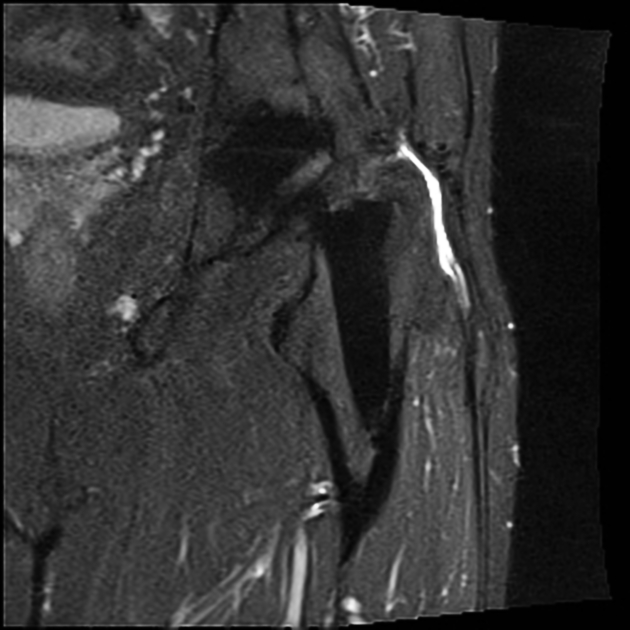
A metal artifact reduction sequence (MARS) is intended to reduce the size and intensity of susceptibility artifacts resulting from magnetic field distortion.
A variety of techniques are used for reducing metal artifacts at MRI, both for addressing artifacts due to the presence of metal in the image plane (in-plane artifacts) and for artifacts due to metal in an adjacent plane (through-plane artifacts).
In-plane artifact reduction
A number of simple changes to the scan protocol can greatly reduce artifacts. Examples are 1:
- lower magnetic field strength: 1.5 T rather than 3 T
- increase bandwidth during slice selection and readout
- increase matrix: 512 pixel
- maintain good signal to noise ratio by increasing number of excitations (NEX)
- spin echo instead of gradient echo where possible
- STIR for fat suppression (spectral frequency selective fat suppression performs better in a homogeneous field)
- shorter echo spacing
- smaller water-fat shift
- thinner slices
- view-angle-tilting (VAT)
Through-plane artifact reduction
Multiacquisition variable-resonance image combination (MAVRIC) is a specialized sequence designed by GE to minimize metallic artifact around metallic prostheses 3. It relies on 3D FSE sequences, using multiple different overlapping volumes at different frequency offsets.
Another technique developed by Siemens used for addressing through-plane metal artifacts is Slice-Encoding for Metal Artifact Correction (SEMAC), where an additional slice-encoding gradient is added to a standard fast-spin echo sequence 4. The combination of the MAVRIC and SEMAC technique is known as MAVRIC-SL 5.
See also
References
- 1. Hargreaves BA, Worters PW, Pauly KB et-al. Metal-induced artifacts in MRI. AJR Am J Roentgenol. 2011;197 (3): 547-55. doi:10.2214/AJR.11.7364 - Pubmed citation
- 2. Hartley KG, Damon BM, Patterson GT et-al. MRI techniques: a review and update for the orthopaedic surgeon. J Am Acad Orthop Surg. 2012;20 (12): 775-87. J Am Acad Orthop Surg (full text) - doi:10.5435/JAAOS-20-12-775 - Pubmed citation
- 3. Hayter CL, Koff MF, Shah P et-al. MRI after arthroplasty: comparison of MAVRIC and conventional fast spin-echo techniques. AJR Am J Roentgenol. 2011;197 (3): W405-11. doi:10.2214/AJR.11.6659 - Pubmed citation
- 4. Sutter R, Ulbrich EJ, Jellus V, Nittka M, Pfirrmann CW. Reduction of metal artifacts in patients with total hip arthroplasty with slice-encoding metal artifact correction and view-angle tilting MR imaging. Radiology. 2012;265(1):204-14. doi:10.1148/radiol.12112408 - Pubmed citation
- 5. Choi SJ, Koch KM, Hargreaves BA, Stevens KJ, Gold GE. Metal artifact reduction with MAVRIC SL at 3-T MRI in patients with hip arthroplasty. AJR Am J Roentgenol. 2015;204(1):140-7. doi:10.2214/AJR.13.11785 - Pubmed citation
Incoming Links
- Metal-on-metal pseudotumour
- Cervical spine protocol (MRI)
- Elbow protocol (MRI)
- MSK pelvis protocol (MRI)
- Thoracic spine protocol (MRI)
- Hip protocol (MRI)
- Reverse total shoulder arthroplasty
- Shoulder protocol (MRI)
- Wrist protocol (MRI)
- Finger protocol (MRI)
- Lumbar spine protocol (MRI)
- Ankle protocol (MRI)
- Medical abbreviations and acronyms (S)
- Medical abbreviations and acronyms (M)
- Reaction to metal
- Mid- and forefoot protocol (MRI)
Related articles: Imaging technology
- imaging technology
- imaging physics
- imaging in practice
-
x-rays
- x-ray physics
- x-ray in practice
- x-ray production
- x-ray tube
- filters
- automatic exposure control (AEC)
- beam collimators
- grids
- air gap technique
- cassette
- intensifying screen
- x-ray film
- image intensifier
- digital radiography
- digital image
- mammography
- x-ray artifacts
- radiation units
- radiation safety
- radiation detectors
- fluoroscopy
-
computed tomography (CT)
- CT physics
- CT in practice
- CT technology
- CT image reconstruction
- CT image quality
- CT dose
-
CT contrast media
-
iodinated contrast media
- agents
- water soluble
- water insoluble
- vicarious contrast material excretion
- iodinated contrast media adverse reactions
- agents
- non-iodinated contrast media
-
iodinated contrast media
-
CT artifacts
- patient-based artifacts
- physics-based artifacts
- hardware-based artifacts
- ring artifact
- tube arcing
- out of field artifact
- air bubble artifact
- helical and multichannel artifacts
- CT safety
- history of CT
-
MRI
- MRI physics
- MRI in practice
- MRI hardware
- signal processing
-
MRI pulse sequences (basics | abbreviations | parameters)
- T1 weighted image
- T2 weighted image
- proton density weighted image
- chemical exchange saturation transfer
- CSF flow studies
- diffusion weighted imaging (DWI)
- echo-planar pulse sequences
- fat-suppressed imaging sequences
- gradient echo sequences
- inversion recovery sequences
- metal artifact reduction sequence (MARS)
-
perfusion-weighted imaging
- techniques
- derived values
- saturation recovery sequences
- spin echo sequences
- spiral pulse sequences
- susceptibility-weighted imaging (SWI)
- T1 rho
- MR angiography (and venography)
-
MR spectroscopy (MRS)
- 2-hydroxyglutarate peak: resonates at 2.25 ppm
- alanine peak: resonates at 1.48 ppm
- choline peak: resonates at 3.2 ppm
- citrate peak: resonates at 2.6 ppm
- creatine peak: resonates at 3.0 ppm
- functional MRI (fMRI)
- gamma-aminobutyric acid (GABA) peak: resonates at 2.2-2.4 ppm
- glutamine-glutamate peak: resonates at 2.2-2.4 ppm
- Hunter's angle
- lactate peak: resonates at 1.3 ppm
- lipids peak: resonates at 1.3 ppm
- myoinositol peak: resonates at 3.5 ppm
- MR fingerprinting
- N-acetylaspartate (NAA) peak: resonates at 2.0 ppm
- propylene glycol peak: resonates at 1.13 ppm
-
MRI artifacts
- MRI hardware and room shielding
- MRI software
- patient and physiologic motion
- tissue heterogeneity and foreign bodies
- Fourier transform and Nyquist sampling theorem
- MRI contrast agents
- MRI safety
-
ultrasound
- ultrasound physics
-
transducers
- linear array
- convex array
- phased array
- frame averaging (frame persistence)
- ultrasound image resolution
- imaging modes and display
- pulse-echo imaging
- real-time imaging
-
Doppler imaging
- Doppler effect
- color Doppler
- power Doppler
- B flow
- color box
- Doppler angle
- pulse repetition frequency and scale
- wall filter
- color write priority
- packet size (dwell time)
- peak systolic velocity
- end-diastolic velocity
- resistive index
- pulsatility index
- Reynolds number
- panoramic imaging
- compound imaging
- harmonic imaging
- elastography
- scanning modes
- 2D ultrasound
- 3D ultrasound
- 4D ultrasound
- M-mode
-
ultrasound artifacts
- acoustic shadowing
- acoustic enhancement
- beam width artifact
- reverberation artifact
- ring down artifact
- mirror image artifact
- side lobe artifact
- speckle artifact
- speed displacement artifact
- refraction artifact
- multipath artifact
- anisotropy
- electrical interference artifact
- hardware-related artifacts
- Doppler artifacts
- aliasing
- tissue vibration
- spectral broadening
- blooming
- motion (flash) artifact
- twinkling artifact
- acoustic streaming
- biological effects of ultrasound
- history of ultrasound
-
nuclear medicine
- nuclear medicine physics
- detectors
- tissue to background ratio
-
radiopharmaceuticals
- fundamentals of radiopharmaceuticals
- radiopharmaceutical labeling
- radiopharmaceutical production
- nuclear reactor produced radionuclides
- cyclotron produced radionuclides
- radiation detection
- dosimetry
- specific agents
- carbon-11
- chromium-51
- fluorine agents
- gallium agents
- Ga-67 citrate
- Ga-68
- iodine agents
-
I-123
- I-123 iodide
- I-123 ioflupane (DaTSCAN)
- I-123 ortho-iodohippurate
- I-131
-
MIBG scans
- I-123 MIBG
- I-131 MIBG
-
I-123
- indium agents
- In-111 Octreoscan
- In-111 OncoScint
- In-111 Prostascint
- In-111 oxine labeled WBC
- krypton-81m
- nitrogen-13
- oxygen-15
- phosphorus-32
- selenium-75
-
technetium agents
- Tc-99m DMSA
- Tc-99m DTPA
- Tc-99m DTPA aerosol
- Tc-99m HMPAO
- Tc-99m HMPAO labeled WBC
- Tc-99m MAA
- Tc-99m MAG3
- Tc-99m MDP
- Tc-99m mercaptoacetyltriglycine
- Tc-99m pertechnetate
- Tc-99m labeled RBC
- Tc-99m sestamibi
- Tc-99m sulfur colloid
- Tc-99m sulfur colloid (oral)
- thallium-201 chloride
- xenon agents
- in vivo therapeutic agents
- pharmaceuticals used in nuclear medicine
-
emerging methods in medical imaging
- radiography
- phase-contrast imaging
- CT
- deep-learning reconstruction
- photon counting CT
- virtual non-contrast imaging
- ultrasound
- magnetomotive ultrasound (MMUS)
- superb microvascular imaging
- ultrafast Doppler imaging
- ultrasound localization microscopy
- MRI
- nuclear medicine
- total body PET system
- immuno-PET
- miscellaneous
- radiography


 Unable to process the form. Check for errors and try again.