
Neurofibroma
Updates to Article Attributes
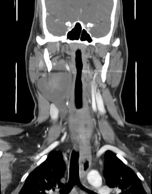
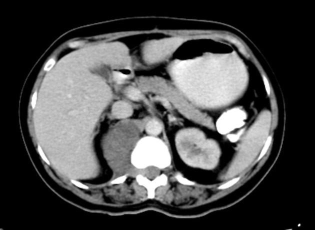
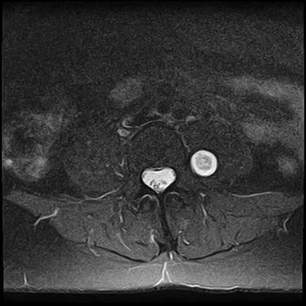
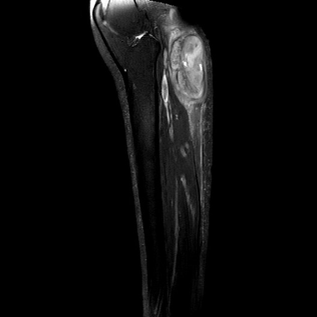
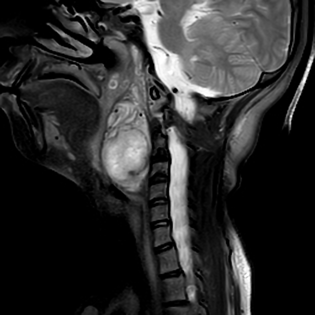
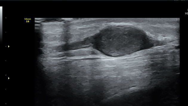
Neurofibromas are benign peripheral nerve sheath tumours usually solitary and sporadic, however, there is a strong association with neurofibromatosis type 1 (NF1). These tumours present as a well-defined hypodense mass with minimal or no contrast enhancement on CT. On MRI, they usually are T1 hypointense and T2 hyperintense with heterogeneous contrast enhancement.
Three types have been describedNeurofibromas are generally divided into three subtypes 1-8:
- localised neurofibroma
- localised cutaneous neurofibroma
- localised intraneural neurofibroma
- diffuse cutaneous neurofibroma (superficial)
- plexiform neurofibroma (deep)
Diffuse cutaneous neurofibromas and plexiform neurofibromas are discussed separately, withand localised cutaneous neurofibromas are generally not a radiological diagnosis. As such, the remainder of this article being a general discussion focusing on the most common localised typeintraneural neurofibromas which isare by far the most common form of neurofibroma, representing 90% of these lesions 2.
Epidemiology
Peak presentation is between 20 and 30 years of age 5 with no sex predilection.
Associations
The majority of localised intraneural neurofibromas are solitary and sporadic and not associated with neurofibromatosis type 1. However, when multiple neurofibromas are present (or plexiform neurofibromas) then the diagnosis of NF1 is almost assured.
Clinical presentation
The clinical presentation of localised intraneural neurofibromas is non-specific and the result of either mass effect on surrounding lesions (or palpable lump) or neurogenic dysfunction.
Pathology
NeurofibromasLocalised intraneural neurofibromas are benign neoplasms composed Schwann cells and fibroblasts, containing a rich network of collagen fibres. They are mostly indolent tumours that rarely only undergo malignant transformation into a malignantperipheral nerve sheath tumour (MPNST); this is only really seen in large neurofibromas, and even then it only occurs in 5-10% of tumours 6.
Neurofibromas are therefore considered WHO grade I tumours under the current (2016) WHO classification of CNS tumours 6.
Macroscopic appearance
Unlike schwannomas, neurofibromas are not encapsulated and infiltrate between the nerve fascicles. They primarily affect superficial cutaneous nerves, however occasionally affect larger deep-seated nerves.
Radiographic features
General imaging features of neurofibromas:
CT
- well-defined hypodense mass
- minimal or no contrast enhancement
MRI
- T1: hypointense
-
T2
- hyperintense
-
target sign
-
- this is thought to be due to a dense central area of collagenous stroma
- although this sign is highly suggestive of neurofibroma, it is occasionally also seen in schwannomas and malignant peripheral nerve sheath tumours
-
- fascicular sign
- T1 C+ (Gd): heterogeneous enhancement
Treatment and prognosis
- lesions not associated with NF1
- localised and diffuse lesions may be treated surgically
- however, as neurofibromas infiltrate between nerve fascicles, they are unable to be separated from the parent nerve and complete excision requires the sacrifice of the nerve
- deep-seated lesions are therefore often managed conservatively
- local recurrence after excision is uncommon and malignant transformation is rare 2
- lesions associated with NF1
- due to the multiplicity of lesions, unless debilitating symptoms are present, treatment of patients with NF1 is often non-surgical
- plexiform neurofibromas are particularly difficult to resect, often leading to incomplete resection.
- recurrence after resection is frequent
- plexiform neurofibromas demonstrate a significant potential for malignant transformation
References changed:
- 8. Mark J. Kransdorf, Mark D. Murphey. Imaging of Soft Tissue Tumors. (2006) ISBN: 9780781747714 - <a href="http://books.google.com/books?vid=ISBN9780781747714">Google Books</a>


 Unable to process the form. Check for errors and try again.