
This is a basic article for medical students and other non-radiologists
Osteoarthritis is a destructive joint pathology that results from longstanding, repetitive trauma. It is most common in the small joints in the hand and weight-bearing joints (knee and hip) and tends to be symmetrical.
On this page:
Terminology
Osteoarthritis (OA) is commonly used in clinical practice and by the public. This common use refers to secondary osteoarthritis. There is also a hereditary form called primary osteoarthritis that occurs primarily in middle-aged women.
Background
Pathology
Secondary osteoarthritis is caused by repeated microtrauma to the joint surface over many years, which results in cartilage thinning and focal reduction in periarticular bone density. These changes result in relative weakness of the bone and overlying cartilage at weight-bearing joints.
Over time, this repeated microtrauma leads to progressive injury, eventually joint space narrowing and destruction of the joint surfaces.
Etiology
A number of risk factors have been found that predispose individuals to the development of OA. These include obesity and metabolic disease, smoking, bone density, and genetic factors 2.
Epidemiology
Secondary osteoarthritis is a relatively common condition and tends to occur in the older population. Women are more commonly affected than men (13% vs 10% 1). Reported incidence is different for hand, knee and hip involvement and varies from country to country 2. The incidence of joint involvement is greatest in the hand, followed by the knee and then the hip.
Clinical features
Presentation
Patients with hip and knee osteoarthritis present with pain and stiffness. They may also have a reduction in range of movement and reduced exercise tolerance.
Diagnosis
Following history taking and examination, plain x-rays are usually all that is required to confirm the diagnosis and make an assessment of severity.
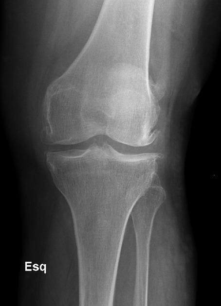
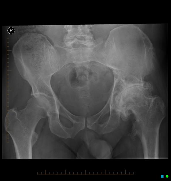
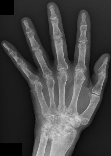
Radiographic features
X-ray
X-rays demonstrate a number of findings in osteoarthritis although they are not always seen and may occur in a variable manner from individual to individual:
joint space narrowing: reflects the loss of cartilage
sclerosis: present in almost all cases of OA
osteophytes: new bone formation around the joint (see osteophytes)
subchondral lucency: focal loss of bone density


 Unable to process the form. Check for errors and try again.