
Pathological fracture
Citation, DOI, disclosures and article data
At the time the article was created Jeremy Jones had no recorded disclosures.
View Jeremy Jones's current disclosuresAt the time the article was last revised Daniel MacManus had no financial relationships to ineligible companies to disclose.
View Daniel MacManus's current disclosures- Pathological fractures
- Pathologic fractures
- Pathologic fracture
- Pathological #
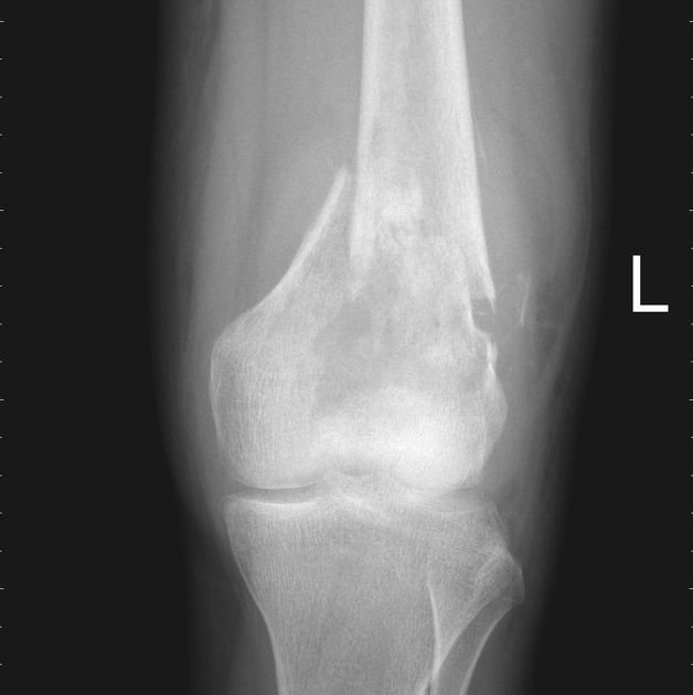
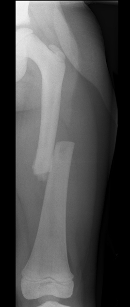
Pathological fractures are fractures that occur in abnormal bone and occur spontaneously or following minor trauma that would not otherwise fracture biomechanically normal bone.
On this page:
Terminology
The term pathological fracture is usually reserved for tumours, either benign or malignant, although it has been used in the setting of osteomyelitis. It can be used in the setting of generalised metabolic bone disease (e.g. Paget disease, osteopetrosis), although the term insufficiency fracture is probably more correct 4. Insufficiency fractures are fractures due to multiple minor events causing a cumulative load on weakened osteoporotic bone. Fragility fractures, on the other hand, are acute fractures in osteoporotic patients due to a single event of minimal trauma.
Pathology
Location
The most common locations for pathological fractures are 4:
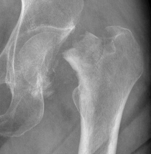
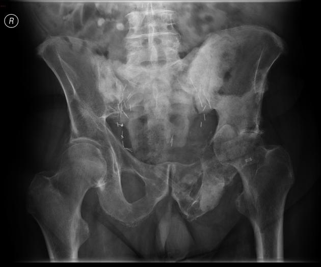
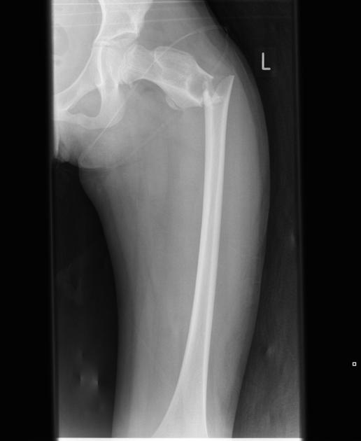
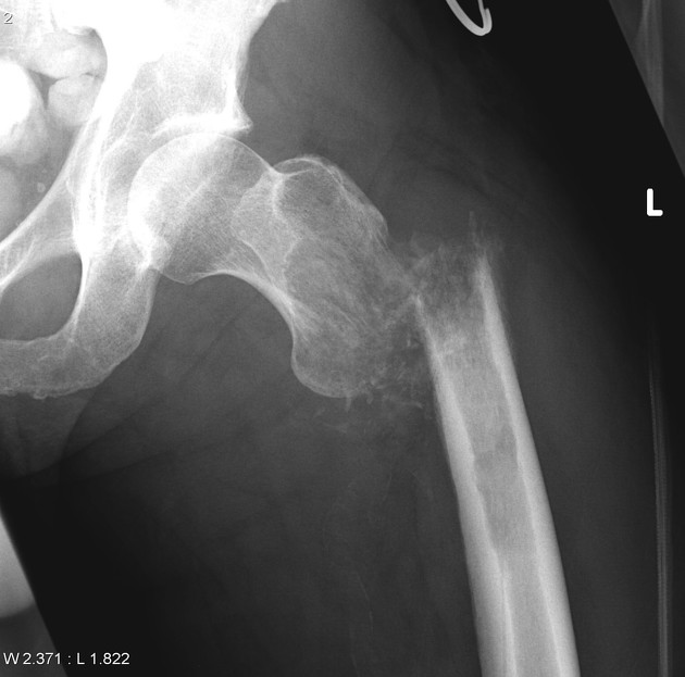
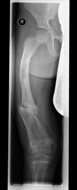
subtrochanteric femur
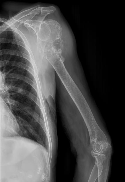
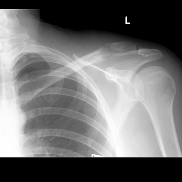
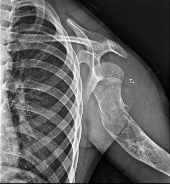
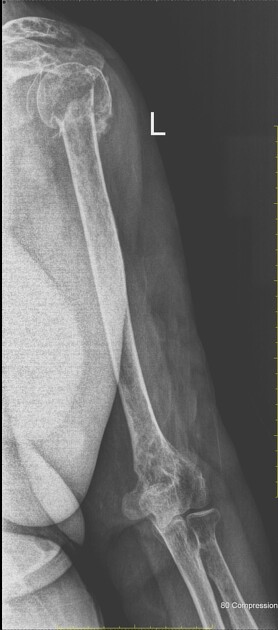
humeral head and metaphyseal junction
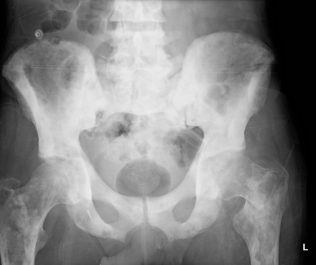
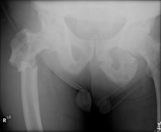
In addition, in adult patients, the avulsion of the femoral lesser trochanter should be considered a pathological fracture until proven otherwise 4.
Radiographic features
Radiography and CT
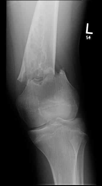
destruction or focal defect within the inner layer of the bony cortex
aggressive periosteal reaction
lytic lesion
soft-tissue mass
mineralised matrix 4
MRI
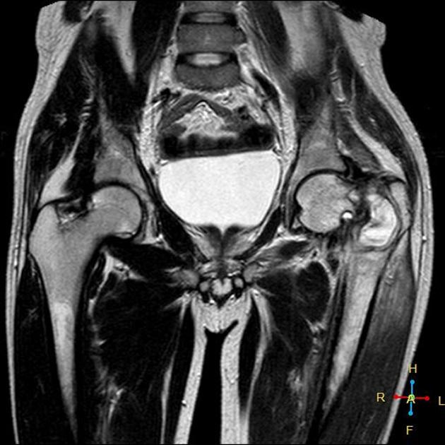
absent or ill-defined fracture line
well-defined, homogenous T1-hypointense abnormal signal without normal intervening marrow signal
adjancent muscle oedema 4
Scintigraphy
diffuse uptake of radiopharmaceutical by the lesion 4
Treatment and prognosis
Pathological fractures are feared by oncologists because they may cause immobilisation of their patients, especially when the spine or lower extremities are affected.
Practical points
A radiologist should mention the possibility of a pathological fracture if an osteolytic metastasis is seen. In principle, every osteolytic focus in the femoral neck or the spine is at risk of a pathological fracture.
Scoring systems have been developed to assess the fracture risk of bone metastases, the Mirels classification is the one that has gained the most traction, although its poor sensitivity (35%) means that it is not without its controversy 5.
Quiz questions
Question 1700
A 45-year-old male who was previously active in sports, now complains of worsening of right hip pain and is unable to bear weight. Pelvic radiograph demonstrates a lucent bone lesion with periosteal reaction at the right lesser trochanter. Bone scan performed with technetium 99m-methyl diphosphonate (99mTc MDP) demonstrates intense tracer uptake in the right subtrochanteric region. No suspicious foci elsewhere. Which diagnosis should be considered until proven otherwise?
References
- 1. Fayad L, Kamel I, Kawamoto S, Bluemke D, Frassica F, Fishman E. Distinguishing Stress Fractures from Pathologic Fractures: A Multimodality Approach. Skeletal Radiol. 2005;34(5):245-59. doi:10.1007/s00256-004-0872-9 - Pubmed
- 2. Fayad L, Kawamoto S, Kamel I et al. Distinction of Long Bone Stress Fractures from Pathologic Fractures on Cross-Sectional Imaging: How Successful Are We? AJR Am J Roentgenol. 2005;185(4):915-24. doi:10.2214/AJR.04.0950 - Pubmed
- 3. Jung H, Jee W, McCauley T, Ha K, Choi K. Discrimination of Metastatic from Acute Osteoporotic Compression Spinal Fractures with MR Imaging. Radiographics. 2003;23(1):179-87. doi:10.1148/rg.231025043 - Pubmed
- 4. Marshall R, Mandell J, Weaver M, Ferrone M, Sodickson A, Khurana B. Imaging Features and Management of Stress, Atypical, and Pathologic Fractures. Radiographics. 2018;38(7):2173-92. doi:10.1148/rg.2018180073 - Pubmed
- 5. Jawad M & Scully S. In Brief: Classifications in Brief: Mirels' Classification: Metastatic Disease in Long Bones and Impending Pathologic Fracture. Clin Orthop Relat Res. 2010;468(10):2825-7. doi:10.1007/s11999-010-1326-4 - Pubmed
- 6. Palmer W, Bancroft L, Bonar F et al. Glossary of Terms for Musculoskeletal Radiology. Skeletal Radiol. 2020;49(Suppl 1):1-33. doi:10.1007/s00256-020-03465-1 - Pubmed
Incoming Links
- Fallen fragment sign
- Femoral diaphyseal stress injury
- Gaucher disease
- Intraosseous schwannoma
- Dedifferentiated chondrosarcoma
- Radius and ulnar shaft fractures
- Fracture
- Distal appendicular bone metastases
- Osteomyelitis
- Enchondroma
- Hypovitaminosis C (scurvy)
- Describing a bone lesion
- Humeral shaft fracture
- Paget disease (bone)
- Kyphosis
- Bone tumors (overview)
- Cloaca (osteomyelitis)
- McCune-Albright syndrome
- Multiple myeloma
- Central atypical cartilaginous tumour/low-grade chondrosarcoma
- Multiple myeloma with pathological fractures
- Polyostotic fibrous dysplasia
- Lytic bone lesions due to multiple myeloma with pathologic intertrochanteric fracture
- Simple bone cyst of the proximal humerus
- Pathologic lesser trochanter avulsion fracture
- Pathologic basicervical hip fracture
- Carbon fiber implant in orthopedic oncology
- Impending pathological fracture of tibia
- Pathological fracture - humerus
- Simple bone cyst - humerus
- Pathological fracture of clavicle
- Cervical spine giant cell tumor
- Odontoid process osteomyelitis with pathological fracture
- Pathological right femur basicervical fracture
- Aneurysmal bone cyst
- Multiple myeloma
- Osteopetrosis
- Unicameral bone cyst with fracture - femur
- Phalangeal enchondroma
- Pathological distal femur fracture
Related articles: Fractures
-
fracture
- terminology
- fracture location[+][+]
- diaphyseal fracture
- metaphyseal fracture
- physeal fracture
- epiphyseal fracture
- fracture types
- avulsion fracture
- articular surface injuries[+][+]
- complete fracture[+][+]
- incomplete fracture[+][+]
- infraction
- compound fracture[+][+]
- pathological fracture
- stress fracture[+][+]
- fracture displacement[+][+]
- fracture location[+][+]
- fracture healing[+][+]
- skull fractures[+][+]
-
facial fractures[+][+]
- fractures involving a single facial buttress
- alveolar process fractures
- frontal sinus fracture
- isolated zygomatic arch fractures
- mandibular fracture
- nasal bone fracture
- orbital blow-out fracture
- paranasal sinus fractures
- complex fractures
- dental fractures
- fractures involving a single facial buttress
-
spinal fractures[+][+]
- classification (AO Spine classification systems)
-
cervical spine fracture classification systems
- AO classification of upper cervical injuries
- AO classification of subaxial injuries
- Anderson and D'Alonzo classification (odontoid fracture)
- Roy-Camille classification (odontoid process fracture)
- Gehweiler classifcation (atlas fractures)
- Levine and Edwards classification (hangman fracture)
- Allen and Ferguson classification (subaxial spine injuries)
- subaxial cervical spine injury classification (SLIC)
- thoracolumbar spinal fracture classification systems
- three column concept of spinal fractures (Denis classification)
- classification of sacral fractures
-
cervical spine fracture classification systems
- spinal fractures by region
- spinal fracture types
- classification (AO Spine classification systems)
- rib fractures[+][+]
- sternal fractures
-
upper limb fractures[+][+]
- classification
- Rockwood classification (acromioclavicular joint injury)
- AO classification (clavicle fracture)
- Neer classification (clavicle fracture)
- Neer classification (proximal humeral fracture)
- AO classification (proximal humeral fracture)
- AO/OTA classification of distal humeral fractures
- Milch classification (lateral humeral condyle fracture)
- Weiss classification (lateral humeral condyle fracture)
- Bado classification of Monteggia fracture-dislocations (radius-ulna)
- Mason classification (radial head fracture)
- Frykman classification (distal radial fracture)
- Mayo classification (scaphoid fracture)
- Hintermann classification (gamekeeper's thumb)
- Eaton classification (volar plate avulsion injury)
- Keifhaber-Stern classification (volar plate avulsion injury)
- upper limb fractures by region
- shoulder
- clavicular fracture
-
scapular fracture
- acromion fracture
- coracoid process fracture
- glenoid fracture
- humeral head fracture
- proximal humeral fracture
- humeral neck fracture
- arm
- elbow
- forearm
- wrist
-
carpal bones
- scaphoid fracture
- lunate fracture
- capitate fracture
- triquetral fracture
- pisiform fracture
- hamate fracture
- trapezoid fracture
- trapezium fracture
- hand
- shoulder
- classification
- lower limb fractures[+][+]
- classification by region
- pelvic fractures
- hip fractures
- Pipkin classification (femoral head fracture)
- Garden classification (hip fracture)
- American Academy of Orthopaedic Surgeons classification (periprosthetic hip fracture)
- Cooke and Newman classification (periprosthetic hip fracture)
- Johansson classification (periprosthetic hip fracture)
- Vancouver classification (periprosthetic hip fracture)
- femoral
- knee
- Schatzker classification (tibial plateau fracture)
- AO classification of distal femur fractures
- Meyers and McKeevers classification (anterior cruciate ligament avulsion fracture)
- tibia/fibula
- Watson-Jones classification (tibial tuberosity avulsion fracture)
- ankle
- foot
- Berndt and Harty classification (osteochondral lesions of the talus)
- Sanders CT classification (calcaneal fracture)
- Hawkins classification (talar neck fracture)
- Myerson classification (Lisfranc injury)
- Nunley-Vertullo classification (Lisfranc injury)
- pelvis and lower limb fractures by region
- pelvic fracture
- sacral fracture
- coccygeal fracture
-
hip
- acetabular fracture
- femoral head fracture
-
femoral neck fracture
- subcapital fracture
- transcervical fracture
- basicervical fracture
-
trochanteric fracture
- pertrochanteric fracture
- intertrochanteric fracture
- subtrochanteric fracture
- femur
- mid-shaft fracture
- bisphosphonate-related fracture
- distal femoral fracture
- knee
- avulsion fractures
- Segond fracture
- reverse Segond fracture
- anterior cruciate ligament avulsion fracture
- posterior cruciate ligament avulsion fracture
- arcuate complex avulsion fracture (arcuate sign)
- biceps femoris avulsion fracture
- iliotibial band avulsion fracture
- semimembranosus tendon avulsion fracture
- Stieda fracture (MCL avulsion fracture)
- patellar fracture
- tibial plateau fracture
- avulsion fractures
- leg
- tibial tuberosity avulsion fracture
- tibial shaft fracture
- fibular shaft fracture
- Maisonneuve fracture
- ankle
- foot
- tarsal bones
- metatarsal bones
- phalanges
- classification by region
- terminology


 Unable to process the form. Check for errors and try again.