
Pericardial calcification is usually seen in individual patients with a history of pericarditis and may be associated with constrictive pericarditis.
On this page:
Pathology
Although historically infective pericarditis was the most common cause, a wide variety of insults can lead to calcification of the pericardium.
- pericarditis: tuberculous, fungal, viral or pyogenic
- previous trauma (hemopericardium)
- cardiac surgery
- collagen vascular diseases: as systemic lupus erythematosus
- uremic pericarditis
- later sequelae of rheumatic heart disease
- malignant pericardial involvement (e.g. mediastinal teratoma)
- post-radiotherapy 1
- idiopathic
- calcified pericardial mass or cyst
Radiographic features
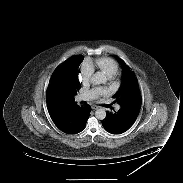
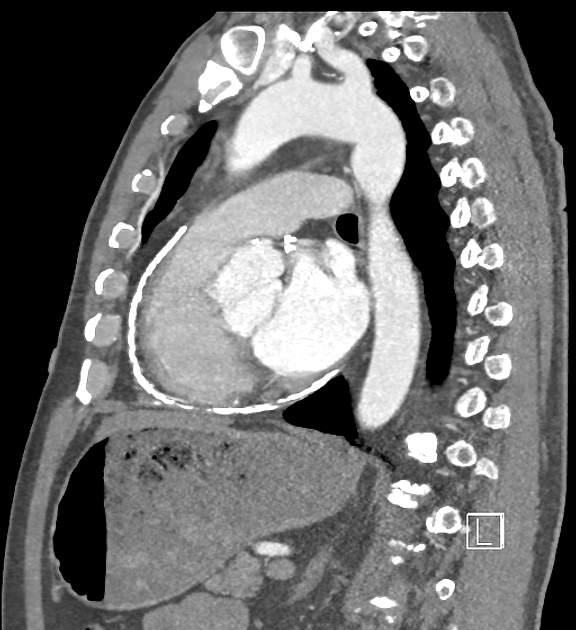
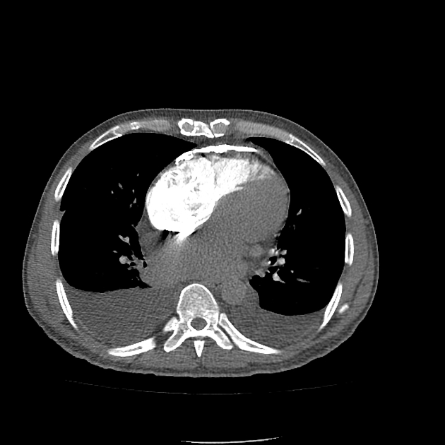
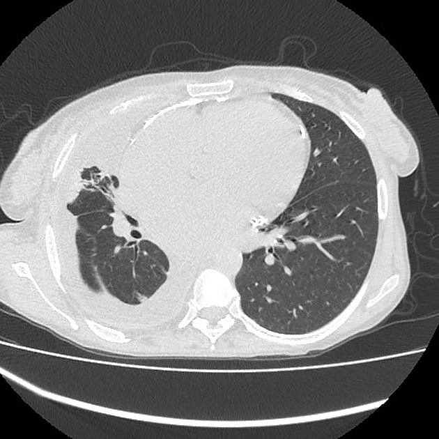
Pericardial calcification is more common over the right side, anterior and diaphragmatic aspects of the heart in the atrioventricular grooves 2. Calcifications over the left ventricle or cardiac apex are rare, unless pericardial calcification is extensive. It is important to assess for signs of associated constrictive pericarditis.
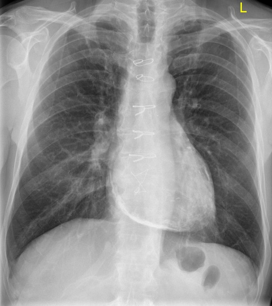
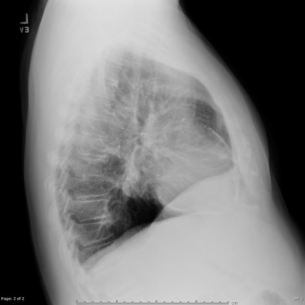
Plain radiograph
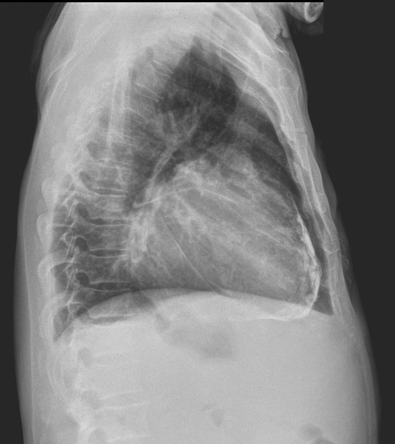
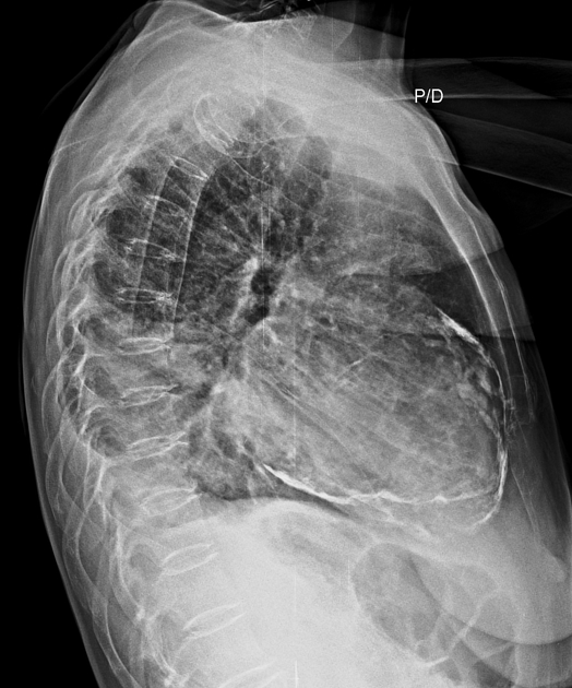
- a curvilinear density at the extreme margin of the cardiac silhouette (better on lateral view)
- extension of calcification over the pulmonary outflow tract (better on lateral view)
- dilated left atrium: due less pericardial investment, even with pericardial constriction, mimicking mitral stenosis
Tuberculous calcifications are the most dense, in the atrioventricular grooves and appear as thick, amorphous oblique circles or arcs of calcifications then spread over the atria and ventricles 2.
Treatment and prognosis
Patients with pericardial calcification may be asymptomatic. If present, symptoms are usually those of heart failure caused by the pericardial calcification restricting the ability of the heart to distend with blood in diastole. In some patients this can be controlled medically, but if the calcification is established this is often ineffective. Pericardiectomy, although not without risk, is potentially curative 3.
Differential diagnosis
The differential diagnosis for pericardial calcifications include:
- constrictive pericarditis: in the setting of heart failure with concern for constrictive pericarditis or restrictive cardiomyopathy, calcifications are highly suggestive of the former
- chronic adhesive pericarditis in the absence of constriction: less dense with a more patchy distribution 4
- rheumatic pericarditis 5
- myocardial calcification: more left-sided and localized calcification (e.g. over the cardiac apex from prior infarction) 6,7


 Unable to process the form. Check for errors and try again.