Phthisis bulbi, also known as end-stage eye, is an atrophic scarred and disorganized non-functioning globe that may result from a variety of severe ocular insults.
On this page:
Epidemiology
In general, phthisis bulbi involves older to elderly patients, usually 65-85 years of age 7. Children and adolescents are only rarely affected, mainly due to ocular trauma, neoplasm and congenital abnormalities 7.
Clinical presentation
Typical clinical symptoms and signs include chronic ocular hypotony, a shrunken globe, pseudoenophthalmos, intraocular tissue fibrosis and scarring, visual loss, recurrent episodes of intraocular irritation, pain and swelling in and around the eye 7,8.
Pathology
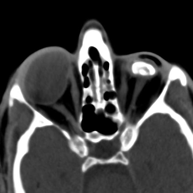
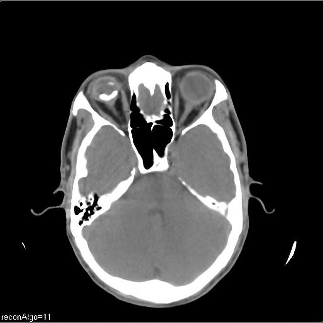
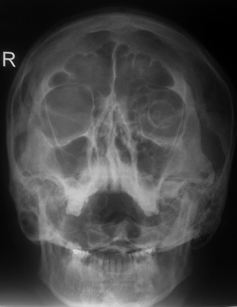
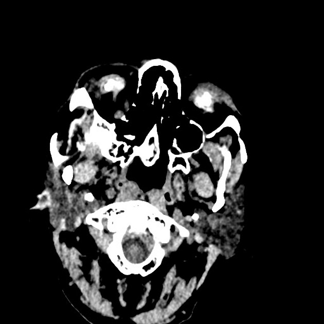
The globe is reduced in size (usually <20 mm) with a thickened/folded posterior sclera. Dystrophic calcification is common, and osseous metaplasia may occasionally occur, forming intraocular bone.
Etiology
Causes of this end-stage damage include:
trauma
infection
other inflammatory processes
radiation
chronic retinal detachment
Radiographic features
CT
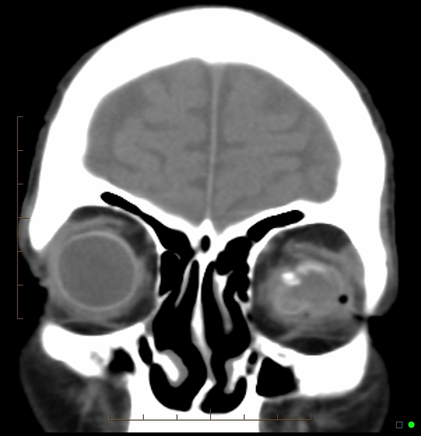
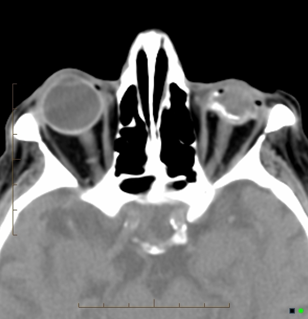
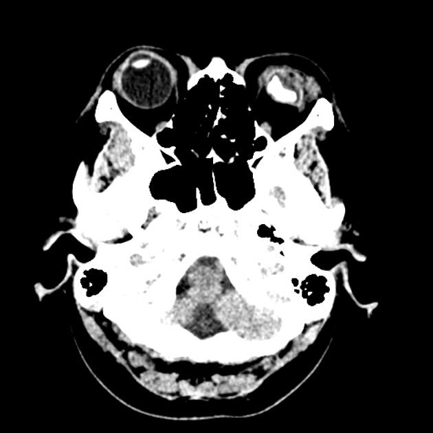
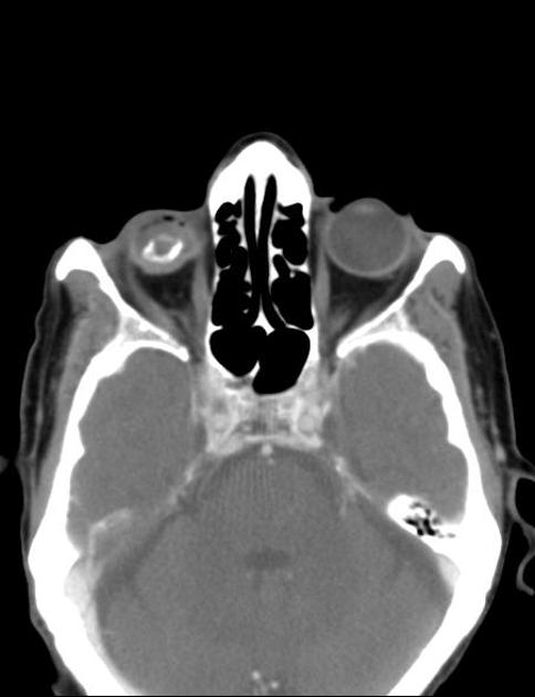
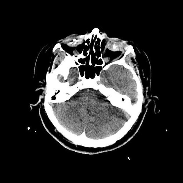
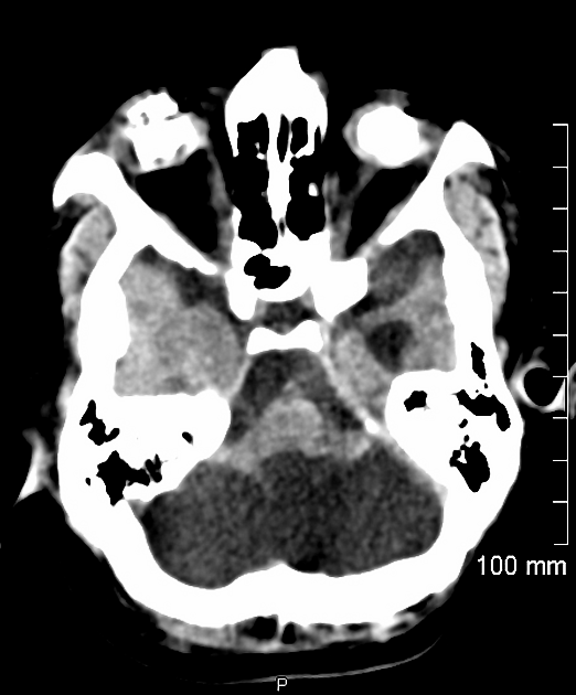
small and shrunken globe with foci of calcium deposits +/- ossification in the sclera, cornea, lens, retina, and optic nerve
distortion of globe components, with loss of the ability to identify separate structures
fibrotic scarring with irregular globe contour and diffusely increased attenuation
MRI
General features:
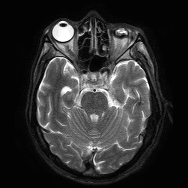
small shrunken, deformed, calcified globe with enophthalmos
abnormal intraocular contents / loss of ability to identify separate structures
Signal characteristics:
T1: often heterogeneous areas of increased signal, depending on the degree of calcification and hemorrhage
T2: heterogeneous vitreous hypointense foci due to coarse calcifications
FLAIR: usually increased signal
Treatment and prognosis
The globe is non-functioning, thus the patient is blind in that eye. Enucleation +/- prosthesis insertion is performed if there is associated chronic pain or for cosmetic reasons.
Differential diagnosis
The differential includes other causes of calcification of the globe.




 Unable to process the form. Check for errors and try again.