
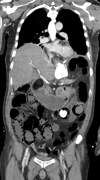
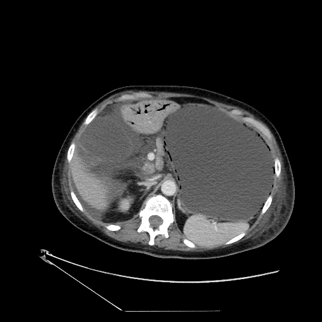
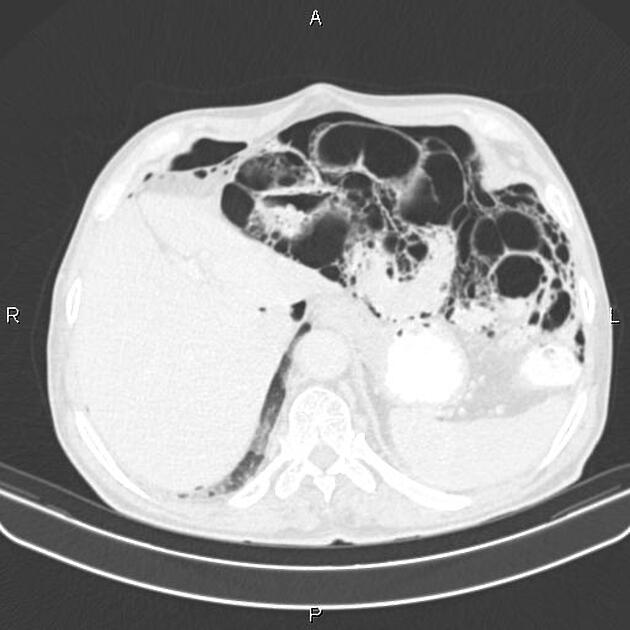
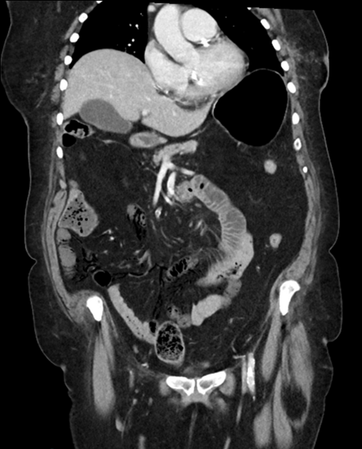
Intramural bowel gas, also known as pneumatosis intestinalis, refers to the clinical or radiological finding of gas within the wall of the bowel (small or large)
On this page:
Terminology
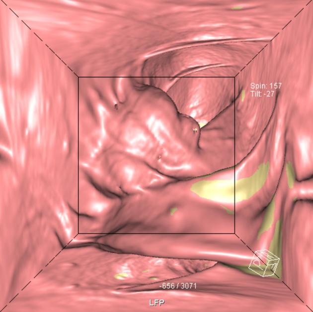
Slightly different sub-terminologies feature in the literature, such as pneumatosis intestinalis (small bowel) and pneumatosis coli (large bowel). Pneumatosis cystoides intestinalis describes the presence of multiple gaseous cysts along the wall of the large bowel, or colon. It is generally an incidental finding in asymptomatic patients.
Pathology
The most common etiology for intramural gas is in intestinal ischemia and bowel infarction, leading to luminal gas penetrating the bowel wall.
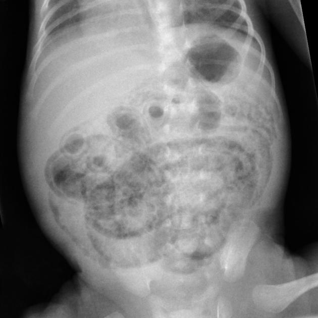
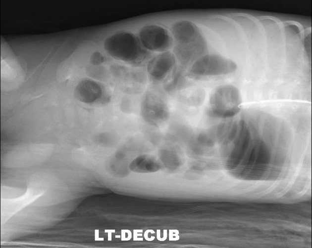
Gas in the bowel wall in the neonatal period, whatever its shape, is diagnostic of necrotizing enterocolitis.
Asymptomatic pneumatosis intestinalis may result from a variety of interrelated contributing factors including:
mucosal integrity
intraluminal pressure
bacterial flora
intraluminal gas
Due to disruption in mucosal integrity with increased mucosal permeability, gas-forming bacteria can enter the submucosa and can produce predominantly hydrogen gas. Another theory is mechanical pressure from pulmonary diseases like COPD leads to pneumatosis intestinalis.
Benign pneumatosis can be caused by a variety of conditions such as pulmonary disease, systemic disease (scleroderma, lupus, AIDS), intestinal inflammation, iatrogenic/procedures, medications (steroids, chemotherapeutic drugs, lactulose, sorbitol and voglibose), and organ transplantation 4.
Life-threatening pneumatosis can be caused by intestinal ischemia, obstruction, enteritis/colitis, toxic caustic ingestion, toxic megacolon, organ transplantation, and collagen vascular disease 4.
Radiographic features
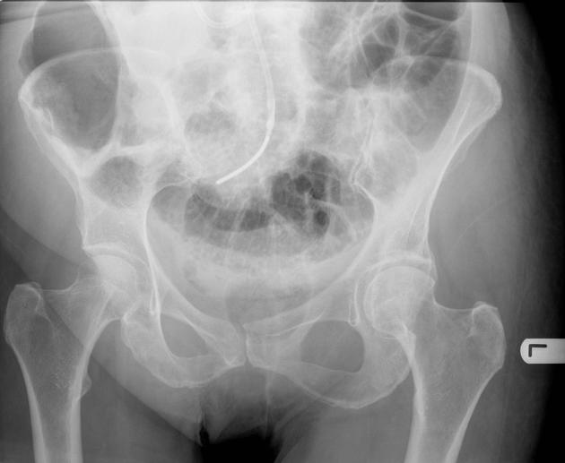
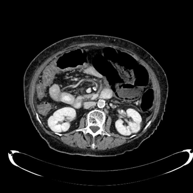
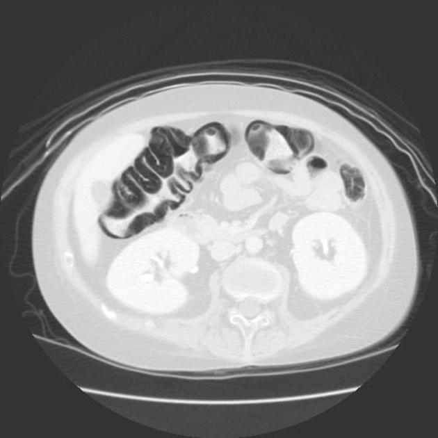
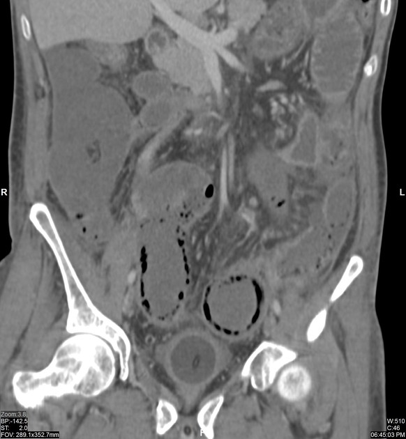
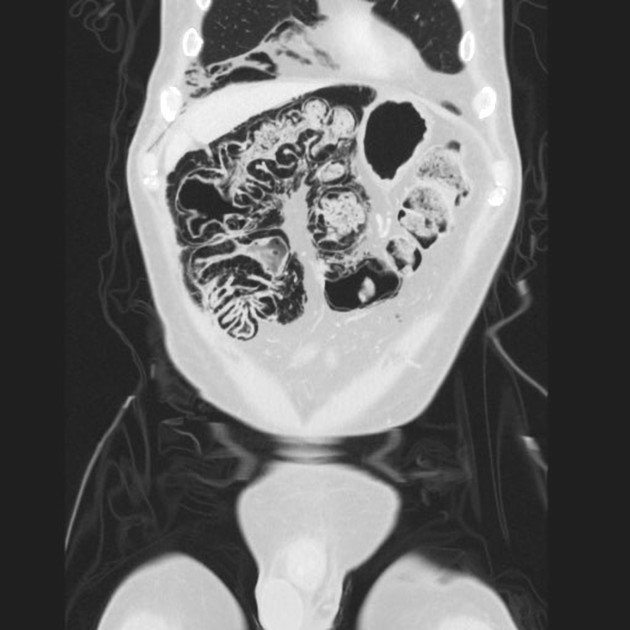
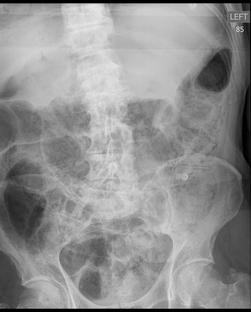
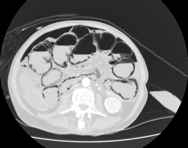
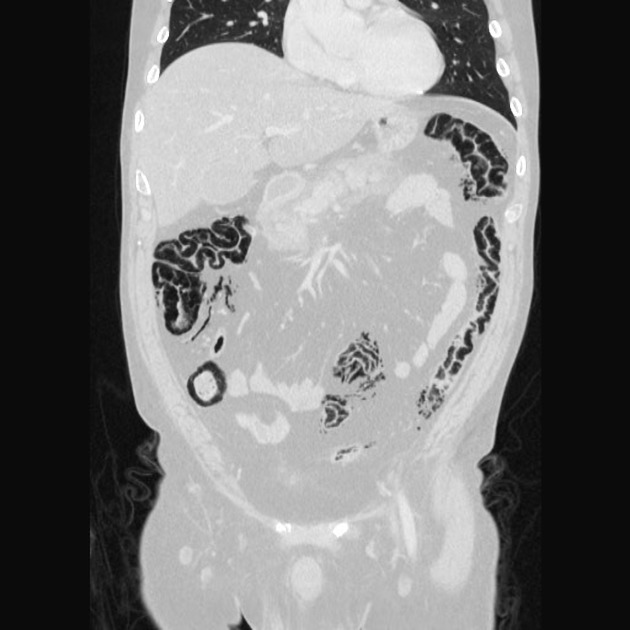
Gas in the bowel wall is most easily identified with CT and plain radiography, but ultrasound and MRI can be useful in pediatric patients to avoid ionizing radiation.
Gas tracks along the bowel wall, appearing as either linear lucencies, which are usually submucosal, or rounded cystic "bubbly" collections, which are usually subserosal 1. Where they join, they may outline the circumferential margin of the bowel, creating rings (this circular pattern of pneumatosis intestinalis favors a benign pathology, whereas the linear and bubbly lucencies can be associated with either benign or life threatening causes).
The following are concerning imaging features of pneumatosis 4,5:
soft tissue bowel wall thickening
free intraperitoneal fluid
lesser extent of pneumatosis (more extensive pneumatosis is more commonly benign)
peri-intestinal soft tissue stranding
abnormal bowel wall enhancement
atherosclerosis and vascular occlusion
The presence of pneumomediastinum favors a benign cause 1. Pneumoperitoneum and pneumoretroperitoneum can be seen with both idiopathic and ischemic pneumatosis 3.
Differential diagnosis
bowel ischemia and infarction
infection
-
medication-induced
chemotherapy
steroid use
autoimmune disease and immunosuppression
connective tissue disorders
-
pulmonary disease
idiopathic
-
iatrogenic
post endoscopy / colonoscopy
post operative
post enteric tube
CT colonography
-
pseudopneumatosis (mimics) include:
gas trapped between bowel wall and luminal contents
gas trapped by opposing mucosal folds
gas bubbles adherent to bowel wall
Practical points
From a clinical perspective, it is essential not to confuse the incidental imaging finding of asymptomatic pneumatosis with symptomatic colonic perforation because the treatment is significantly different 2.


 Unable to process the form. Check for errors and try again.