
Pulmonary lymphangiectasia (PL) refers to a rare, fatal congenital abnormality of the lungs characterized by grossly dilated lymphatic channels in the sub pleural, interlobar, perivascular and peribronchial areas 3.
It is divided into two main types 1:
cardiac-associated lymphangiectasia (secondary type)
-
non-cardiac associated lymphangiectasia (primary type)
early-onset type
late-onset type
On this page:
Clinical presentation
Clinical features depend on the type:
-
cardiac-associated lymphangiectasia (secondary type)
cyanosis and respiratory depression
-
non-cardiac associated lymphangiectasia (primary type)
-
early-onset type
abnormal pattern of respiration
-
late-onset type
progressive pattern of lung disease starting at 10 days old 1
-
Pathology
Abnormal development of the lungs and associated lymphatics during 14th to 20th week of gestation 1.
During the intrauterine life, in this phase of gestation, the lymphatics in the sub pleural space and interlobular areas are large and decrease in caliber by the 20th week. However, continued development of the lung without decrease in size of lymphatics results in this abnormality.
Associations
-
cardiac-associated lymphangiectasia (secondary type)
-
there is variable configuration of the heart depending on the underlying cardiac malformation; most commonly:
TAPVR to SVC, innominate vein, ductus venosus and portal vein
-
-
non-cardiac associated lymphangiectasia (primary type)
-
early-onset type
congenital ichthyosis
urethral stenosis
-
late-onset type
obstructive uropathy
-
Radiographic features
Ultrasound
hydrops fetalis (associated feature)
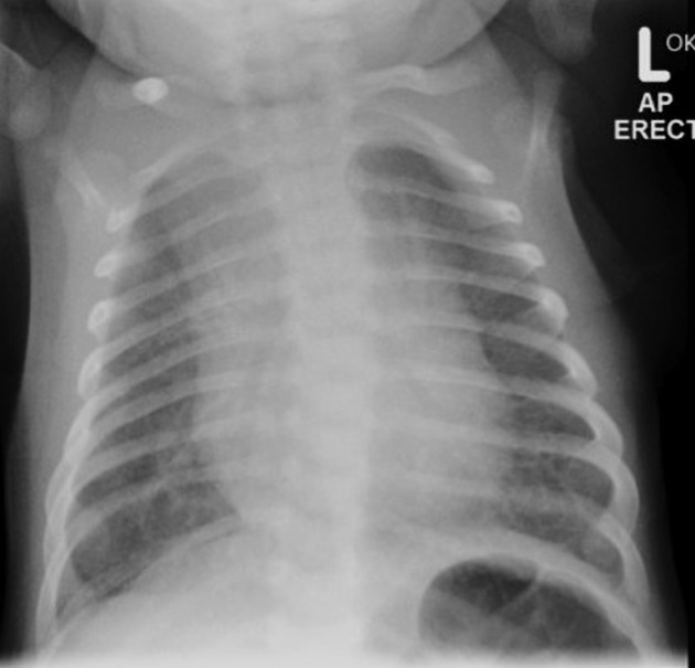
Plain radiograph
perihilar infiltrates
pleural effusion
varying degrees of hyperinflation
HRCT
perihilar infiltrates with air bronchograms
interstitial and interlobular septal thickening
pleural effusion (chylothorax)
MRI
-
T1 (especially coronal):
interstitial thickening
pleural effusion
atelectasis
-
T2 (especially axial)
usually shows high-signal material within the pulmonary interstitium 3
Lymphoscintigraphy
radiotracer accumulation in the lungs
asymmetric visualization of lymphatic channels
Treatment and prognosis
Cardiac associated and the non-cardiac associated early onset types are more fatal leading to death. The late onset type does not have a poor prognosis as symptoms and clinical findings improve after the first year of life 5.


 Unable to process the form. Check for errors and try again.