
The radial bands sign, also known as radial migration bands, refers to linear bands seen on MRI, radiating from the periventricular white matter to the subcortical region, thought to be specific for tuberous sclerosis 1,2.
On this page:
Pathology
The exact pathogenesis of radial bands is uncertain, but they are thought to relate to dysfunction of, or injury to, the radial glial fibers (which go on to transform into astrocytes) and form the scaffolding over which neurons migrate from the periventricular germinal matrix to the cortex 2,3.
Radiographic features
MRI
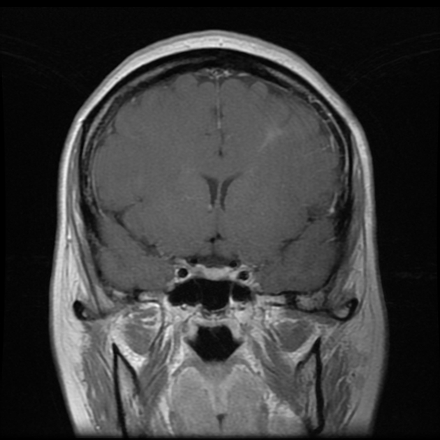
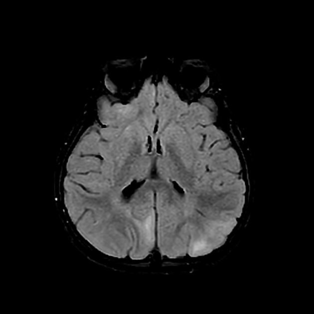
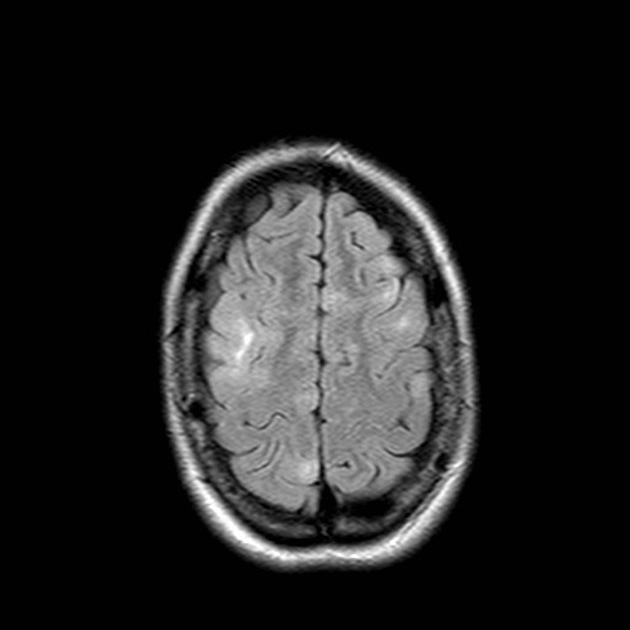
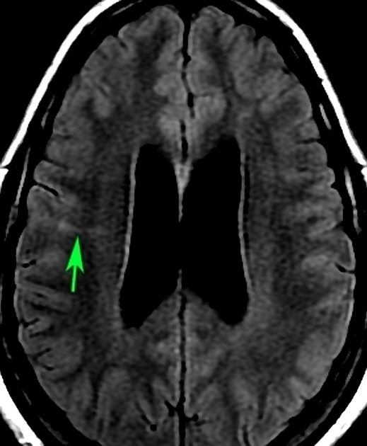
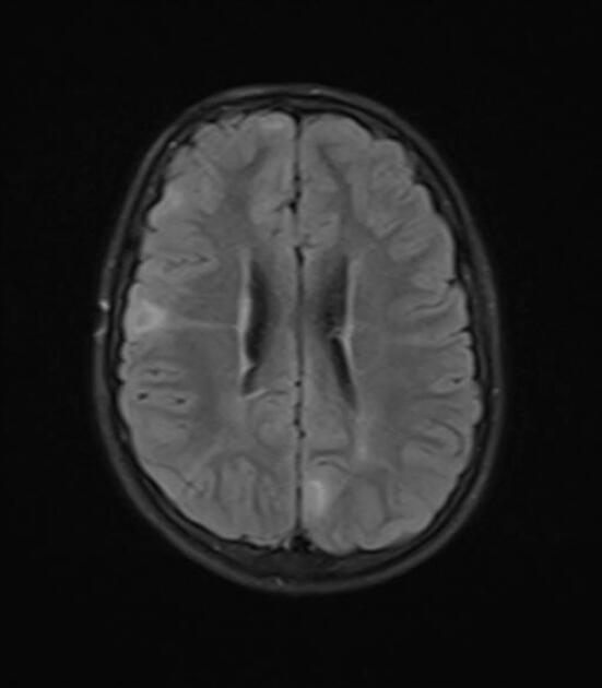
These radial bands appear as linear regions of signal abnormality extending from the ventricle to the cortex, slightly fanning out as they reach the periphery.
Signal characteristics in adults are 1,2:
T1: iso to hypointense
T2/FLAIR: hyperintense
T1 C+ (Gd): occasional enhancement is encountered
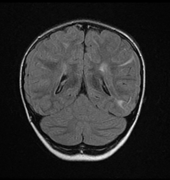
In young patients with incomplete myelination, signal characteristics are 2:
T1: hyperintense to unmyelinated white matter
T2: iso to hypointense
Differential diagnosis
The main differential is with the transmantle sign of type II focal cortical dysplasia. Indeed, whether there is a meaningful difference between radial band sign and transmantle sign is debatable and the two may represent the same histopathological abnormality 4.


 Unable to process the form. Check for errors and try again.