
Sublingual gland
Citation, DOI, disclosures and article data
At the time the article was created Jeremy Jones had no recorded disclosures.
View Jeremy Jones's current disclosuresAt the time the article was last revised Craig Hacking had the following disclosures:
- Philips Australia, Paid speaker at Philips Spectral CT events (ongoing)
These were assessed during peer review and were determined to not be relevant to the changes that were made.
View Craig Hacking's current disclosures- Sublingual glands
- Sublingual salivary gland
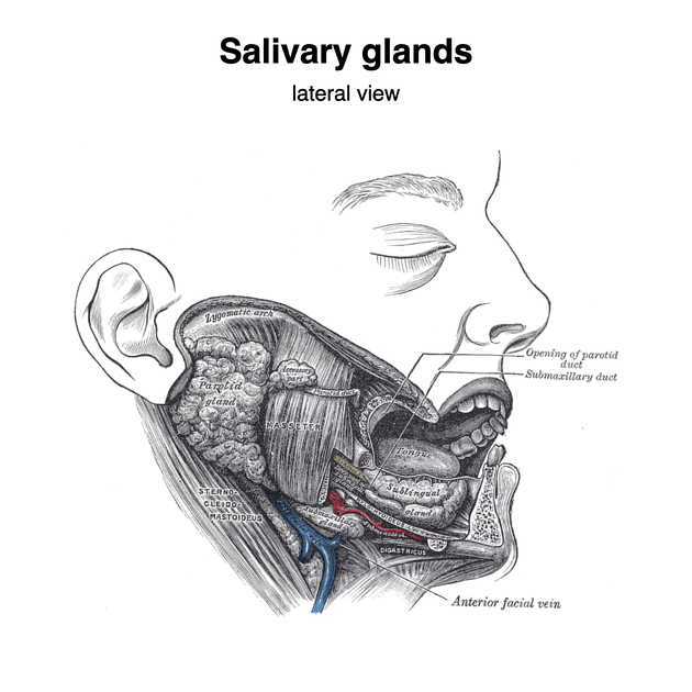
The sublingual glands are the smallest of the three paired major salivary glands that lie in the sublingual space on the floor of the mouth, anterior to the submandibular glands. They secrete predominantly mucous saliva that is drained by numerous ducts, collectively termed the minor sublingual ducts (of Rivinus). Occasionally, the anteriormost duct is enlarged, forming the major sublingual duct (of Bartholin) which opens together with the submandibular duct at the sublingual papilla.
On this page:
Gross anatomy
The sublingual glands are small, almond-shaped glands. They lie against the medial surface of the mandible at the sublingual fossa and are immediately lateral to the submandibular duct and lingual nerve.
The superior surface of the gland indents the mucosa of the floor of the mouth, creating the sublingual fold, which extends to the sublingual papilla at the base of the frenulum of the tongue.
It is the only major salivary gland that does not contain a capsule 5. The parotid and submandibular glands both have a fibrous capsule derived from the superficial (investing) layer of the deep cervical fascia 4.
ADVERTISEMENT: Supporters see fewer/no ads
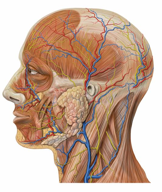
Arterial supply
sublingual artery (branch of the lingual artery)
submental artery (branch of the facial artery)
Venous drainage
sublingual vein (tributary of the lingual vein)
submental vein (tributary of the facial vein)
Innervation
Autonomic innervation is from the lingual nerve via the submandibular ganglion with parasympathetic fibers arising from the chorda tympani and sympathetic fibers from the superior cervical ganglion.
History and etymology
Bartholin duct is named after its discoverer, the Danish anatomist Caspar Bartholin the Younger (1655-1738) 3.
Related pathology
References
- 1. Keith L. Moore, Arthur F. Dalley, A. M. R. Agur. Clinically Oriented Anatomy. (2013) ISBN: 9781451119459 - Google Books
- 2. Last, R. J., McMinn, R. M. H.. Last's Anatomy, Regional and Applied. (1994) ISBN: 044304662X - Google Books
- 3. Hill R. The Contributions of the Bartholin Family to the Study and Practice of Clinical Anatomy. Clin Anat. 2007;20(2):113-5. doi:10.1002/ca.20355 - Pubmed
- 4. Richard Drake, A. Wayne Vogl, Adam W. M. Mitchell. Gray's Anatomy for Students E-Book. (2019) ISBN: 9780323611053 - Google Books
- 5. Grewal J, Bordoni B, Shah J, Ryan J. Anatomy, Head and Neck, Sublingual Gland. 2023. - Pubmed
Incoming Links
- Ranula
- Superior cervical ganglion
- Parasympathetic nervous system
- Chorda tympani
- Salivary gland trauma
- Salivary gland tumours
- Mandible
- Salivary glands
- Lingual vein
- Major salivary glands
- Inferior salivatory nucleus
- Sympathetic nervous system
- Duct of Rivinus
- Superior salivary nucleus
- Major salivary gland cancer (staging)
- Facial nerve
- Major sublingual ducts
- Submandibular ganglion
- Ducts of the salivary glands
- Langerhans cell histiocytosis (salivary gland manifestations)
Related articles: Anatomy: Head and neck
- skeleton of the head and neck
-
cranial vault
- scalp (mnemonic)
- fontanelle
-
sutures
- calvarial
- facial
- frontozygomatic suture
- frontomaxillary suture
- frontolacrimal suture
- frontonasal suture
- temporozygomatic suture
- zygomaticomaxillary suture
- parietotemporal suture (parietomastoid suture)
- occipitotemporal suture (occipitomastoid suture)
- sphenofrontal suture
- sphenozygomatic suture
- spheno-occipital suture (not a true suture)
- lacrimomaxillary suture
- nasomaxillary suture
- internasal suture
- basal/internal
- skull landmarks
- frontal bone
- temporal bone
- parietal bone
- occipital bone
- skull base (foramina)
-
facial bones
- midline single bones
- paired bilateral bones
- cervical spine
- hyoid bone
- laryngeal cartilages
-
cranial vault
- muscles of the head and neck
- muscles of the tongue (mnemonic)
- muscles of mastication
-
facial muscles
- epicranius muscle
- circumorbital and palpebral muscles
- nasal muscles
-
buccolabial muscles
- elevators, retractors and evertors of the upper lip
- levator labii superioris alaeque nasalis muscle
- levator labii superioris muscle
- zygomaticus major muscle
- zygomaticus minor muscle
- levator anguli oris muscle
- malaris muscle
- risorius muscle
- depressors, retractors and evertors of the lower lip
- depressor labii inferioris muscle
- depressor anguli oris muscle
- mentalis muscle
- compound sphincter
-
orbicularis oris muscle
- incisivus labii superioris muscle
- incisivus labii inferioris muscle
-
orbicularis oris muscle
- muscle of mastication
- modiolus
- elevators, retractors and evertors of the upper lip
- muscles of the middle ear
- orbital muscles
- muscles of the soft palate
- pharyngeal muscles
- suprahyoid muscles
- infrahyoid muscles
- intrinsic muscles of the larynx
- muscles of the neck
- platysma muscle
- longus colli muscle
- longus capitis muscle
- scalenus anterior muscle
- scalenus medius muscle
- scalenus posterior muscle
- scalenus pleuralis muscle
- sternocleidomastoid muscle
-
suboccipital muscles
- rectus capitis posterior major muscle
- rectus capitis posterior minor muscle
- obliquus capitis superior muscle
- obliquus capitis inferior muscle
- accessory muscles of the neck
- deep cervical fascia
-
deep spaces of the neck
- anterior cervical space
- buccal space
- carotid space
- danger space
- deep cervical fascia
- infratemporal fossa
- masticator space
- parapharyngeal space
- stylomandibular tunnel
- parotid space
- pharyngeal (superficial) mucosal space
- perivertebral space
- posterior cervical space
- pterygopalatine fossa
- retropharyngeal space
- suprasternal space (of Burns)
- visceral space
- surgical triangles of the neck
- orbit
- ear
- paranasal sinuses
- upper respiratory tract
- viscera of the neck
- blood supply of the head and neck
-
arterial supply
-
common carotid artery
- carotid body
- carotid bifurcation
- subclavian artery
- variants
-
common carotid artery
- venous drainage
-
arterial supply
- innervation of the head and neck
-
cranial nerves
- olfactory nerve (CN I)
- optic nerve (CN II)
- oculomotor nerve (CN III)
- trochlear nerve (CN IV)
-
trigeminal nerve (CN V) (mnemonic)
- trigeminal ganglion
- ophthalmic division
- maxillary division
- mandibular division
- abducens nerve (CN VI)
- facial nerve (CN VII)
-
vestibulocochlear nerve (CN VIII)
- vestibular ganglion (Scarpa's ganglion)
- glossopharyngeal nerve (CN IX)
- vagus nerve (CN X)
- (spinal) accessory nerve (CN XI)
- hypoglossal nerve (CN XII)
- parasympathetic ganglia of the head and neck
- cervical sympathetic ganglia
- greater occipital nerve
- third occipital nerve
-
cervical plexus
- muscular branches
- longus capitis
- longus colli
- scalenes
- geniohyoid
- thyrohyoid
-
ansa cervicalis
- omohyoid (superior and inferior bellies separately)
- sternothyroid
- sternohyoid
- phrenic nerve
- contribution to the accessory nerve (CN XI)
- cutaneous branches
- muscular branches
- brachial plexus
- pharyngeal plexus
-
cranial nerves
- lymphatic drainage of the head and neck
- embryological development of the head and neck


 Unable to process the form. Check for errors and try again.