
Substantia nigra
Citation, DOI, disclosures and article data
At the time the article was created Jeremy Jones had no recorded disclosures.
View Jeremy Jones's current disclosuresAt the time the article was last revised Francis Deng had no financial relationships to ineligible companies to disclose.
View Francis Deng's current disclosures- Substantiae nigrae
- Substantia nigras
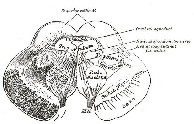
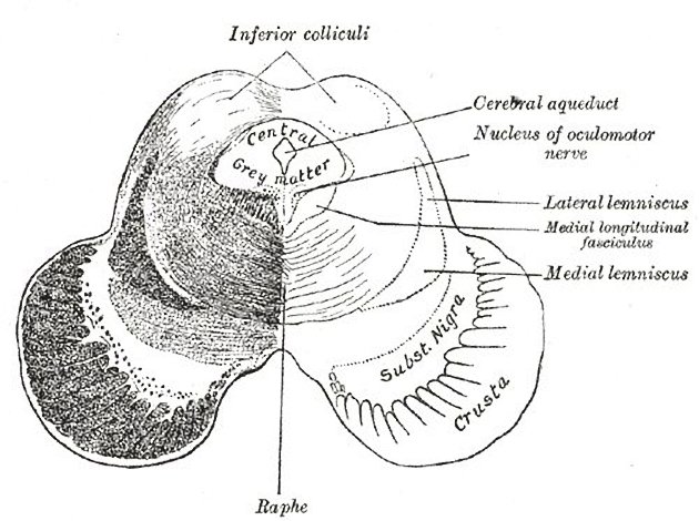
The substantia nigra (plural: substantiae nigrae) is one of the brainstem nuclei and part of the extrapyramidal system. While other nuclei such as the red nucleus are small and contained within an axial slice at the superior colliculi (see figure), the substantia nigra is seen in axial slices at both superior and inferior colliculi.
On this page:
Gross anatomy
They are situated in the anterior midbrain and mark the transition point of the tegmentum and cerebral peduncles.
Function
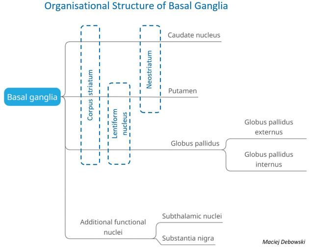
It is an important relay station in the motor system. It consists of a compact part (dark, containing melanin) and a reticular part (reddish, containing iron). Most of its axons are projected diffusely to other brain areas and not arranged into tracts. Numerous axonal tracts terminate in the substantia nigra:
- caudate nucleus (striatonigral fibers)
- anterior cerebral cortex (corticonigral fibers)
- putamen
- precentral cortex
Nigrosomes
Nigrosomes are small clusters of dopaminergic cells measuring up to a few millimeters in size within the substantia nigra that exhibit calbindin D28K negativity on immunohistochemistry. Five nigrasomes have been described with the largest labeled as nigrosome-1 positioned in the caudal, medio-lateral portion of the substantia nigra. Nigrasome-1 contains the largest proportion of neurons affected in Parkinson disease 3.
Radiographic features
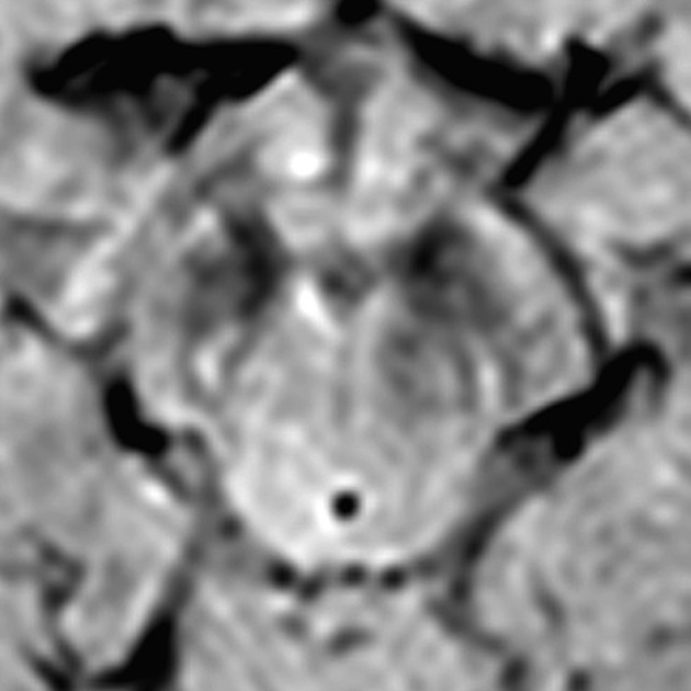
High resolution T2*/SWI weighted MRI is able to identify the distribution of normal signal within healthy and diseased substantia nigra both at 7 T and 3 T field strength. The healthy nigrosome-1 produces a swallow tail appearance on axial imaging. Absence of the normal swallow tail appearance of nigrosome-1 is reported to be a reliable sign of Parkinson disease 3.
Related pathology
References
- 1. Standring S (editor). Gray's Anatomy (39th edition). Churchill Livingstone. (2011) ISBN:0443066841. Read it at Google Books - Find it at Amazon
- 2. Ross LMMP. Atlas of anatomy. George Thieme Verlag. (2007) ISBN:3131421215. Read it at Google Books - Find it at Amazon
- 3. Schwarz ST, Afzal M, Morgan PS et-al. The 'swallow tail' appearance of the healthy nigrosome - a new accurate test of Parkinson's disease: a case-control and retrospective cross-sectional MRI study at 3T. PLoS ONE. 2014;9 (4): e93814. doi:10.1371/journal.pone.0093814 - Free text at pubmed - Pubmed citation
Incoming Links
- McLeod syndrome
- Chorea-acanthocytosis
- Face of the giant panda sign (midbrain)
- Parkinson disease
- Nigrosomes
- Swallow tail sign (substantia nigra)
- Putamen
- Brainstem nuclei
- Cerebral atrophy
- Weber syndrome
- Pantothenate kinase-associated neurodegeneration
- Tegmentum
- Habenula
- Saint Louis encephalitis
- Ventral tegmental area
- Neuroferritinopathy
- Phospholipase A2 associated neurodegeneration
- Oculomotor nerve
- Corpus striatum
- Perry syndrome
Related articles: Anatomy: Brain
-
brain
- grey matter
- white matter
-
cerebrum
-
cerebral hemisphere (telencephalon)
- cerebral lobes and gyri
- frontal lobe
- parietal lobe
-
occipital lobe
- occipital pole
- lingual gyrus
- fusiform gyrus (Brodmann area 37)
- calcarine (visual) cortex
- cuneus
- temporal lobe
- basal forebrain
- limbic system
- insula
-
cerebral sulci and fissures (A-Z)
- calcarine fissure
- callosal sulcus
- central (Rolandic) sulcus
- cingulate sulcus
- collateral sulcus
- inferior frontal sulcus
- inferior occipital sulcus
- inferior temporal sulcus
- interhemispheric fissure
- intraparietal sulcus
- lateral (Sylvian) sulcus
- lateral occipital sulcus
- marginal sulcus
- occipitotemporal sulcus
- olfactory sulcus
- paracentral sulcus
- paraolfactory sulcus
- parieto-occipital fissure
- posterior parolfactory sulcus
- precentral sulcus
- preoccipital notch
- postcentral sulcus
- rhinal sulcus
- rostral sulcus
- subparietal sulcus
- superior frontal sulcus
- superior occipital sulcus
- superior temporal sulcus
- cortical histology
- cerebral lobes and gyri
- white matter tracts
- deep grey matter
-
pituitary gland
- posterior pituitary and stalk (part of diencephalon)
- anterior pituitary
- inferior hypophyseal arterial circle
- diencephalon
-
cerebral hemisphere (telencephalon)
-
brainstem
- midbrain (mesencephalon)
- pons (part of metencephalon)
- medulla oblongata (myelencephalon)
- white matter
-
grey matter
- non-cranial nerve
-
cranial nerve nuclei
- oculomotor nucleus
- Edinger-Westphal nucleus
- trochlear nucleus
- motor nucleus of CN V
- mesencephalic nucleus of CN V
- main sensory nucleus of CN V
- spinal nucleus of CN V
- abducent nucleus
- facial nucleus
- superior salivatory nucleus
- cochlear nuclei
- vestibular nuclei
- inferior salivatory nucleus
- solitary tract nucleus
- ambiguus nucleus
- dorsal vagal motor nucleus
- hypoglossal nucleus
-
cerebellum (part of metencephalon)
- vermis
- cerebellar hemisphere
- cerebellar peduncles
- cranial meninges (meninx primitiva)
- CSF spaces
-
cranial nerves (mnemonic)
- olfactory nerve (CN I)
- optic nerve (CN II)
- oculomotor nerve (CN III)
- trochlear nerve (CN IV)
- trigeminal nerve (CN V) (mnemonic)
- abducens nerve (CN VI)
- facial nerve (CN VII) (segments mnemonic | branches mnemonic)
-
vestibulocochlear nerve (CN VIII)
- vestibular ganglion (Scarpa's ganglion)
- glossopharyngeal nerve (CN IX)
- vagus nerve (CN X)
- spinal accessory nerve (CN XI)
- hypoglossal nerve (CN XII)
- functional neuroanatomy
- CNS development
- cerebral vascular supply
- arteries
- vascular territories
-
circle of Willis
- internal carotid artery (ICA) (segments)
- vertebral artery
-
normal variants
- intracranial arterial fenestration
- internal carotid artery (ICA)
- anterior cerebral artery (ACA)
- middle cerebral artery (MCA)
- posterior cerebral artery (PCA)
- basilar artery
- persistent carotid-vertebrobasilar artery anastomoses (mnemonic)
- vertebral artery
- ophthalmic artery
-
cerebral venous system
-
dural venous sinuses
- basilar venous plexus
- cavernous sinus (mnemonic)
- clival diploic veins
- inferior petro-occipital vein
- inferior petrosal sinus
- inferior sagittal sinus
- intercavernous sinus
- internal carotid artery venous plexus of Rektorzik
- jugular bulb
- marginal sinus
- occipital sinus
- sigmoid sinus
- sphenoparietal sinus
- straight sinus
- superior petrosal sinus
- superior sagittal sinus
- torcula herophili
- transverse sinus
-
cerebral veins
-
superficial veins of the brain
- superior cerebral veins (superficial cerebral veins)
- inferior cerebral veins
- superficial middle cerebral vein
- superior anastomotic vein (of Trolard)
- inferior anastomotic vein (of Labbe)
-
superficial veins of the brain
-
deep veins of the brain
- great cerebral vein (of Galen)
- venous circle of Trolard
- normal variants
-
dural venous sinuses
- arteries
- glymphatic pathway


 Unable to process the form. Check for errors and try again.