
Gastrointestinal manifestations in patients with systemic lupus erythematosus (SLE) are common and may involve any region of the gastrointestinal tract or visceral organs.
On this page:
Clinical presentation
Patients with abdominal or gastrointestinal involvement by systemic lupus erythematosus may have a variety of presentations, including abdominal pain, diarrhea, dysphagia, anorexia, ascites, oral ulcers, gastrointestinal bleeding, and malnutrition depending on the organs involved and duration of involvement ref.
Pathology
SLE may affect any part of the gastrointestinal tract and visceral organs:
peritoneum: ascites in up to 10% of patients due to serositis ref
-
gastrointestinal vasculature
vasculitis causing colitis, mucosal ulceration with hemorrhage and/or perforation, mesenteric ischemia, "watermelon stomach" or gastric antral vascular ectasia, esophageal dysmotility or intestinal pseudo-obstruction 4
thrombosis of the intestinal vessels may also occur in association with antiphospholipid syndrome ref
-
mucosa
mouth ulcers in 50% of patients ref
protein-losing enteropathy and fat malabsorption ref
associated ulcerative colitis or Crohn disease ref
pancreas: pancreatitis
-
liver and biliary tract
hepatomegaly, steatosis, hepatitis, cholestasis, primary biliary cholangitis, and cirrhosis ref
Budd-Chiari syndrome: may be associated with antiphospholipid antibodies in SLE ref
acalculous cholecystitis, benign biliary stricture ref
spleen: splenic infarction ref
adrenal glands: adrenal hematoma, hypoadrenalism ref
Radiographic features
Fluoroscopy
Barium study
small bowel series: prominent mucosal pattern because of edema, segments of spiculation, fragmentation and clumping of barium 2
upper GI series: decreased esophageal peristalsis, reflux, esophageal dilatation ref
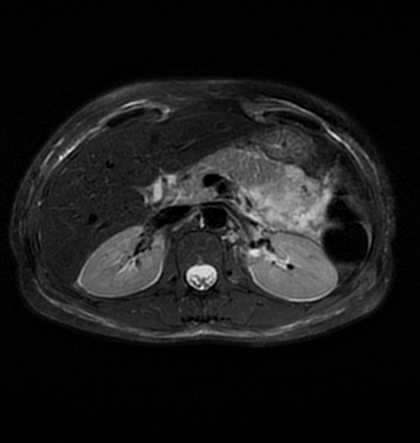
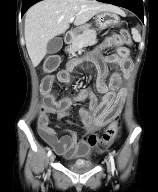
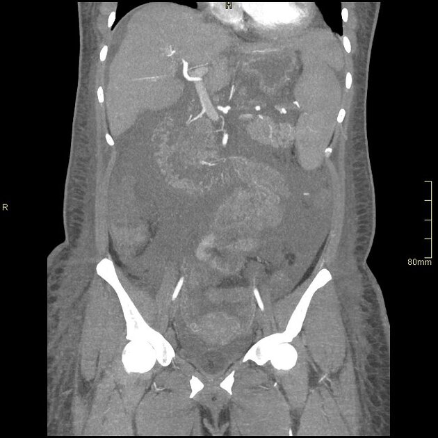
CT
CT findings may include ref:
ascites with increased peritoneal enhancement
circumferential, symmetrical, multisegmented mural thickening usually of the jejunum and ileum with associated submucosal edema (target sign)
engorgement of mesenteric vessels with a palisade pattern or comb-like appearance
dilatation of intestinal segments
mesenteric fat stranding
diffuse splenic calcification in the typical "onion-skin" pattern due to concentric perivascular lamination of fibrous tissue
other features and complications of SLE disease may be present (e.g. pancreatitis, intestinal ischemia)


 Unable to process the form. Check for errors and try again.