
Tracheal bronchus
Citation, DOI, disclosures and article data
At the time the article was created Yuranga Weerakkody had no recorded disclosures.
View Yuranga Weerakkody's current disclosuresAt the time the article was last revised Mostafa Elfeky had no financial relationships to ineligible companies to disclose.
View Mostafa Elfeky's current disclosures- Pig bronchus
- Tracheal bronchi
- Bronchus suis
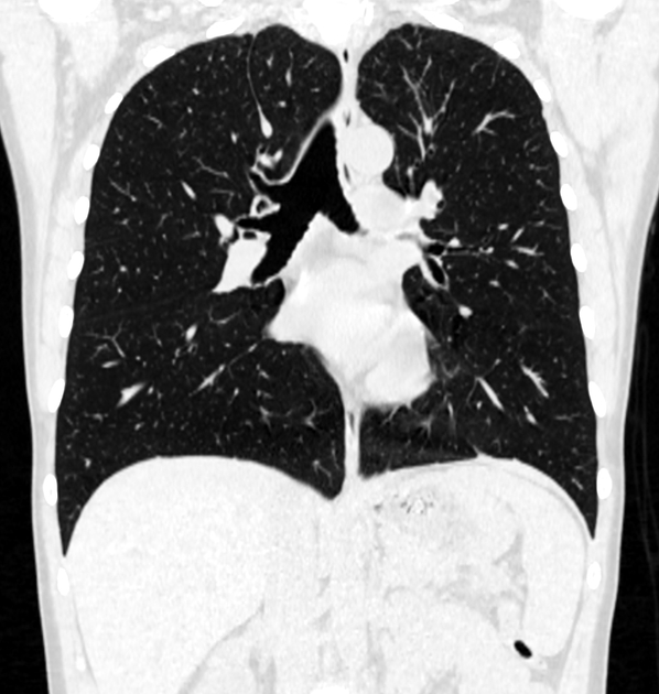
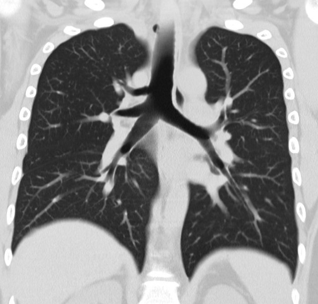
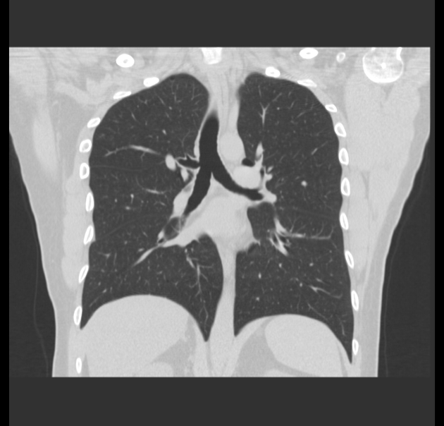
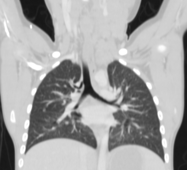
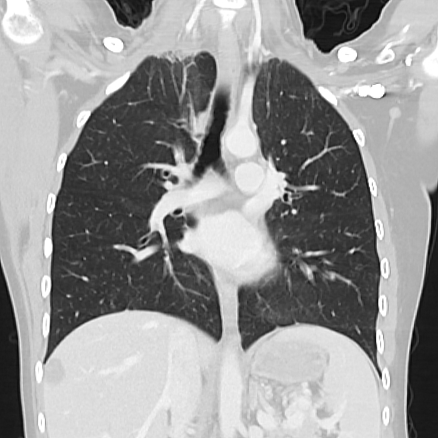
A tracheal bronchus (with some variations also known as a pig bronchus) is an anatomical variant where an accessory bronchus originates directly from the supracarinal trachea.
On this page:
Terminology
The term (pig bronchus or bronchus suis) is often given when the entire upper lobe (usually right side) is supplied by this bronchus 5.
However, this term is used in some literature to encompass a wider spectrum of abnormalities including accessory bronchi originating from either the trachea or main bronchi.
Epidemiology
Incidence is estimated at ~1% (range 0.1-2%), and there is a marked right sided predilection 1,2,5.
Clinical presentation
Often incidentally discovered and most patients are asymptomatic. Occasionally patients may have a recurrent (right) upper lobe pneumonia due to focal emphysematous change.
Pathology
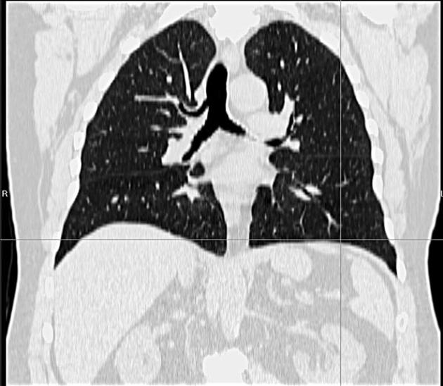
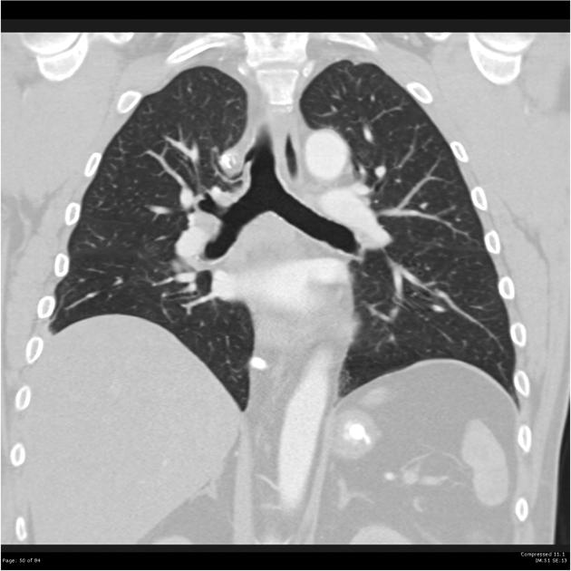
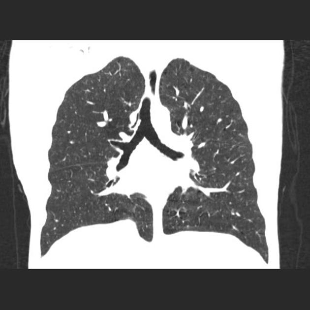
Tracheal bronchi arise from the right lateral wall of the trachea usually at a distance of <2 cm from the level of the carina 5.
They can be classified into two main types:
supernumerary: usual bronchial supply to affected lung segment is concurrently present
displaced: usual bronchial supply to affected lung segment is concurrently absent
Radiographic features
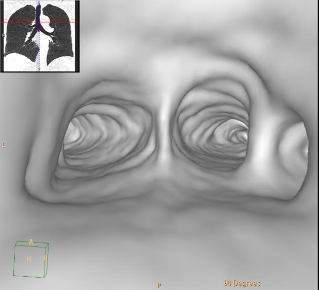
CT
CT is the best modality for assessing the anatomy and allows direct visualisation and orientation of the anomalous bronchus. Coronal multi-planar reconstructions in "lung window" settings are the most helpful and is best in depicting this anomaly.
History and etymology
It was initially described by Sandifort in 1785 2.
References
- 1. Shih FC, Lee WJ, Lin HJ. Tracheal bronchus. CMAJ. 2009;180 (7): 783. doi:10.1503/cmaj.080280 - Free text at pubmed - Pubmed citation
- 2. Ghaye B, Szapiro D, Fanchamps JM et-al. Congenital bronchial abnormalities revisited. Radiographics. 21 (1): 105-19. Radiographics (full text) - Pubmed citation
- 3. Harris JH. Clinical significance of tracheal bronchus. AJR Am J Roentgenol. 1983;141 (3): 623. AJR Am J Roentgenol (citation) - Pubmed citation
- 4. Gower WA, Mcgrath-morrow SA, Macdonald KD et-al. Tracheal bronchus in a 6-month-old infant identified by CT with three-dimensional airway reconstruction. Thorax. 2008;63 (1): 93-4. doi:10.1136/thx.2006.071100 - Pubmed citation
- 5. Müller NL, Silva CI. Imaging of the chest. (2008) ISBN:141604048X. Read it at Google Books - Find it at Amazon
- 6. Han J, Xiang H, Ridley W, Ridley L. Pig Bronchus. J Med Imaging Radiat Oncol. 2018;62:34. doi:10.1111/1754-9485.21_12785
Incoming Links
- Bronchus suis
- Chronic pneumonitis due to displaced segmental bronchus with accessory fissure
- Bronchus suis
- Endotracheal tube obstructing pig bronchus
- Tracheal bronchus
- Tracheal bronchus
- Tetralogy of Fallot
- Tracheal bronchus
- Bronchus suis
- Tracheal bronchus and azygos lobe
- Complete tracheal ring
- Tracheal bronchus - bronchus suis
- Tracheal bronchus
- Tracheal bronchus
- Bronchus suis
- Tracheal bronchus
Related articles: Anatomy: Thoracic
- thoracic skeleton
- thoracic cage
- thoracic spine
- articulations
- muscles of the thorax
- diaphragm
- intercostal space
- intercostal muscles
- variant anatomy
- spaces of the thorax
- thoracic viscera
- lower respiratory tract
-
heart
- cardiac chambers
- heart valves
- cardiac fibrous skeleton
- innervation of the heart
- development of the heart
- cardiac wall
-
pericardium
- epicardium
- epicardial fat pad
- pericardial space
- oblique pericardial sinus
- transverse pericardial sinus
-
pericardial recesses
- aortic recesses
- pulmonic recesses
- postcaval recess
- pulmonary venous recesses
- pericardial ligaments
- myocardium
- endocardium
-
pericardium
- oesophagus
- thymus
- breast
- arterial supply of the thorax
-
thoracic aorta (development)
-
ascending aorta
-
aortic root
- aortic annulus
-
coronary arteries
- coronary arterial dominance
- myocardial segments
-
left main coronary artery (LMCA)
- ramus intermedius artery (RI)
-
circumflex artery (LCx)
- obtuse marginal branches (OM1, OM2, etc))
- Kugel's artery
-
left anterior descending artery (LAD)
- diagonal branches (D1, D2, etc)
- septal perforators (S1, S2, etc)
-
right coronary artery (RCA)
- conus artery
- sinoatrial nodal artery
- acute marginal branches (AM1, AM2, etc)
- inferior interventricular artery (PDA)
- posterior left ventricular artery (PLV)
- congenital anomalies
- sinotubular junction
-
aortic root
- aortic arch
- aortic isthmus
- descending aorta
-
ascending aorta
- pulmonary trunk
-
thoracic aorta (development)
- venous drainage of the thorax
- superior vena cava (SVC)
- inferior vena cava (IVC)
-
coronary veins
-
cardiac veins which drain into the coronary sinus
- great cardiac vein
- middle cardiac vein
- small cardiac vein
- posterior vein of the left ventricle
- vein of Marshall (oblique vein of the left atrium)
- anterior cardiac veins
- venae cordis minimae (smallest cardiac veins or thebesian veins)
-
cardiac veins which drain into the coronary sinus
- pulmonary veins
- bronchial veins
- thoracoepigastric vein
- lymphatics of the thorax
- innervation of the thorax


 Unable to process the form. Check for errors and try again.