
Vestibulocochlear nerve
Updates to Article Attributes
The vestibulocochlear nerve is the eigth (CN VIII) iscranial nerve. It exits the eighth cranialbrainstem through the cerebellopontine angle, passing into the internal acoustic meatus as part of the acousticofacial bundle. Within the internal acoustic meatus, the nerve branches into cochlear and has two roles:
-
innervationvestibular nerves to supply the cochlea, and vestibule, respectively. From these special sensory organs of the internal ear, the vestibulocochlear nerve transmits afferent impulses to thecochleafor hearing -
innervation to thevestibulefor accelerationcentral nervous system encoding auditory stimuli, static (gravitational) equilibrium, andbalance senses
Gross anatomy
NucleiNucleus and intraparenchymal portion
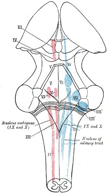
ThereThe dorsal and ventral cochlear nuclei are two special sensory located in the dorsolateral upper medulla, deep to the lateral angle of the rhomboid fossa. Situated medial to the cochlear nuclei are the four vestibular nuclei, which form two columns extending rostrally into the lower pons 1,2. Fibres from the vestibular and four special sensory cochlear nuclei pass around the inferior cerebellar peduncle, exiting the brainstem through the cerebellopontine angle at the pontomedullary junction in the form of distinct vestibular nuclei locatedand cochlear roots 2.
Cisternal portion
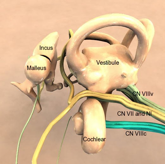
In the cerebellopontine angle, the two roots quickly converge to form a single vestibulocochlear nerve. The nerve makes up the most lateral component of the acousticofacial bundle, the other components of which include the facial nerve, nervus intermedius and labyrinthine artery. The vestibulocochlear nerve passes laterally and slightly anterosuperiorly through the cerebellopontine angle cistern, superior to the petro-occipital fissure, inferior petrosal sinus and posterior aspect of the petrous temporal bone 3. It exits the posterior cranial fossa as part of the acousticofacial bundle, passing through the internal acoustic meatus 1,3.
Intrameatal portion
Travelling laterally within the lower pons and upper medulla.
Vestibulocochlear nerve
It emerges between the pons and the medulla, lateral to the facial nerve and nervus intermedius, passing laterally through the cerebellopontine angle to the internal acoustic meatus (IAM) with, the aforementioned two other nerves.
In the IAM thevestibulocochlear nerve splitsdivides again into four bundles:vestibular and cochlear nerveroots, superior and inferior divisionwhich further divide to supply a number of the vestibular nerve and nerve from the posterior semicircular canal, separated from each other by the falciform crest and Bill bar
Branches
Cochlear nerve
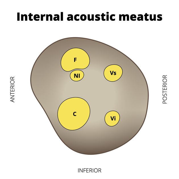
The cochlear nerve relays withpasses through the first-order sensory cellsanteroinferior quadrant of the fundus (most lateral part) of the internal acoustic meatus. It spirals towards the spiral ganglion located in the spiral ganglion, which is in the basespiral lamina
Vestibular nerve
TheAt the fundus of the internal acoustic meatus, the vestibular nerve relays inexpands to form the vestibular ganglion (a.k.a. ganglion of (of Scarpa), from there three from which the following bundles emerge2,3:
-
Thesuperior division, located(utriculo-ampullary nerve): courses in the posterosuperior quadrant of the internalauditory canalacoustic meatus,carriescarrying sensory fibres from the utricle and hair cells of the superior and lateral semicircular canalsandutricle -
Theinferior division, located(saccular nerve): courses in the posteroinferior quadrant of the internal acoustic meatus to enter the vestibule,carriescarrying sensory fibres from the saccule -
Thesingular nerve (posterior ampullary nerve): courses in the posteroinferior quadrant of the internal acoustic meatus and through the foramen singulare, carrying sensory fibres from the posterior semicircular canal, also in the posteroinferior quadrant, passes through theforamen singulare
Related pathology
References changed:
- 1. Keith L. Moore, Arthur F. Dalley, A. M. R. Agur. Clinically Oriented Anatomy. (2013) ISBN: 9781451119459 - <a href="http://books.google.com/books?vid=ISBN9781451119459">Google Books</a>
- 2. Stanley Jacobson, Elliott M. Marcus. Neuroanatomy for the Neuroscientist. (2011) ISBN: 9781441996527 - <a href="http://books.google.com/books?vid=ISBN9781441996527">Google Books</a>
- 3. J. P. Barral, Alain Croibier. Manual Therapy for the Cranial Nerves. (2009) ISBN: 9780702031007 - <a href="http://books.google.com/books?vid=ISBN9780702031007">Google Books</a>
- 4. Chummy S. Sinnatamby. Last's Anatomy. (2011) ISBN: 9780702033940 - <a href="http://books.google.com/books?vid=ISBN9780702033940">Google Books</a>
- 5. Derald E. Brackmann, Clough Shelton, Moises A. Arriaga. Otologic Surgery. (2010) ISBN: 9781416046653 - <a href="http://books.google.com/books?vid=ISBN9781416046653">Google Books</a>
- 1. Last's Anatomy. Churchill Livingstone. (2011) ISBN:0702033952. <a href="http://books.google.com/books?vid=ISBN0702033952">Read it at Google Books</a> - <a href="http://www.amazon.com/gp/product/0702033952">Find it at Amazon</a><span class="ref_v3"></span>
- 2. Clemente CD. Anatomy. Lippincott Williams & Wilkins. (2011) ISBN:1582558892. <a href="http://books.google.com/books?vid=ISBN1582558892">Read it at Google Books</a> - <a href="http://www.amazon.com/gp/product/1582558892">Find it at Amazon</a><span class="ref_v3"></span>


 Unable to process the form. Check for errors and try again.