
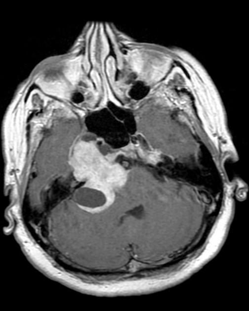
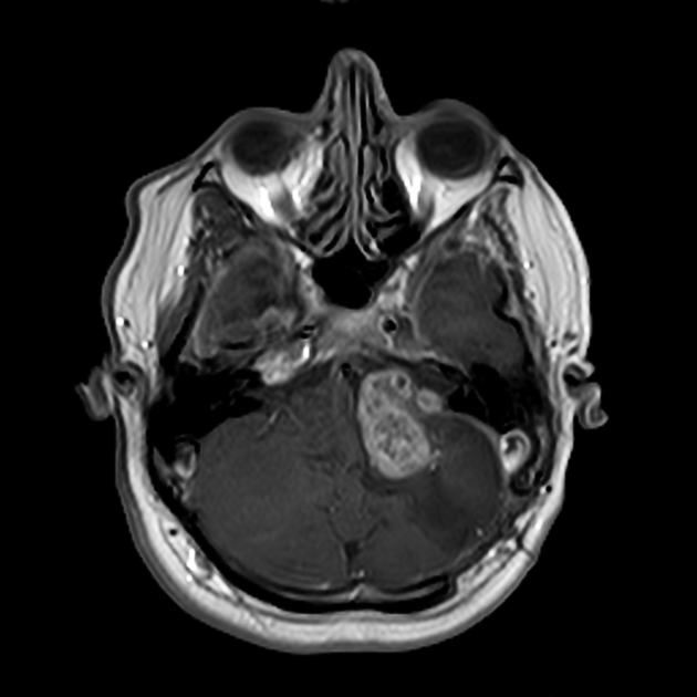
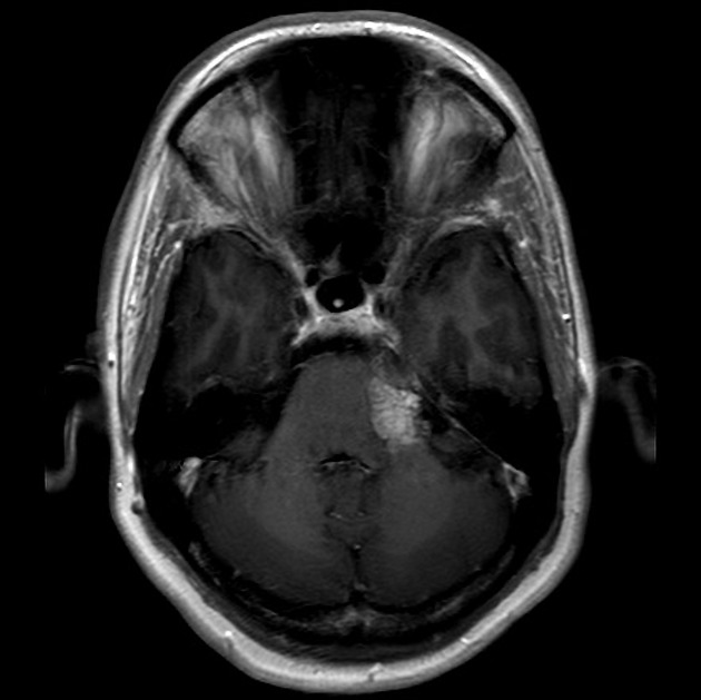
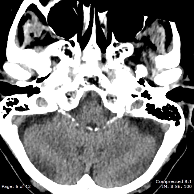
Cerebellopontine angle (CPA) masses are relatively common. Although a diverse range of pathologies may be seen in this region, the most common by far is vestibular schwannoma.
Pathology
Cerebellopontine angle masses can be divided into four groups, based on imaging characteristics:
enhancing mass
mass with high T1 signal on MRI
mass with CSF intensity/density
other masses
Alternatively, a quick mnemonic to remember the common entities affecting the cerebellopontine angle is AMEN or SAME.
Enhancing mass
vestibular schwannoma: most common by far (~80%)
meningioma: second most common (~10%)
metastasis, e.g. breast, lung, malignant melanoma
High T1 signal mass
hemorrhagic vestibular schwannoma
neurenteric cyst: usually prepontine, but fluid may be proteinaceous and high on T1
thrombosed berry aneurysm: often will have a calcified rim and hemosiderin staining
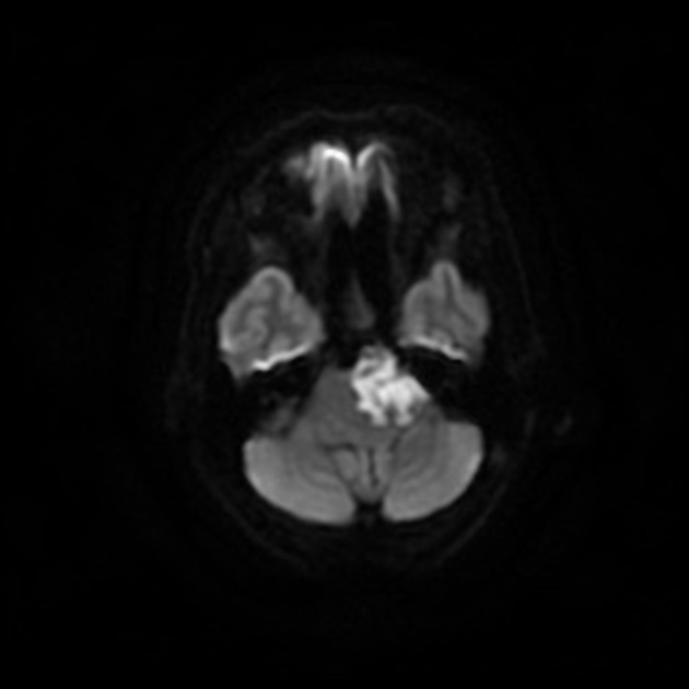
white epidermoid: rare; restricts on DWI
cerebellopontine angle lipoma: usually has the facial nerve and vestibulocochlear nerve coursing through it; saturates on fat suppressed sequences
ruptured intracranial dermoid cyst: often multiple droplets with original midline lesion still often seen
CSF density mass
epidermoid cyst: third most common (~5%)
Other masses
Many other masses can present at or around the cerebellopontine angle. They include:
ganglioglioma (rare 3)
-
mass mimics
calcified choroid plexus from the fourth ventricle protruding through the lateral foramen of Luschke: Bochdalek's flower basket


 Unable to process the form. Check for errors and try again.