
Internal acoustic canal
Citation, DOI, disclosures and article data
At the time the article was created Jeremy Jones had no recorded disclosures.
View Jeremy Jones's current disclosuresAt the time the article was last revised Frank Gaillard had no financial relationships to ineligible companies to disclose.
View Frank Gaillard's current disclosures- Internal acoustic meatus (IAM)
- Internal auditory meatus (IAM)
- Internal auditory canal (IAC)
- Internal acoustic canal (IAC)
- Internal acoustic meatus
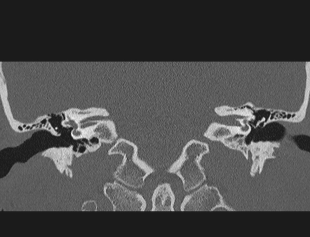
The internal acoustic canal (IAC), also known as the internal auditory canal or meatus (IAM), is a bony canal within the petrous portion of the temporal bone that transmits nerves and vessels from within the posterior cranial fossa to the auditory and vestibular apparatus.
On this page:
Gross anatomy
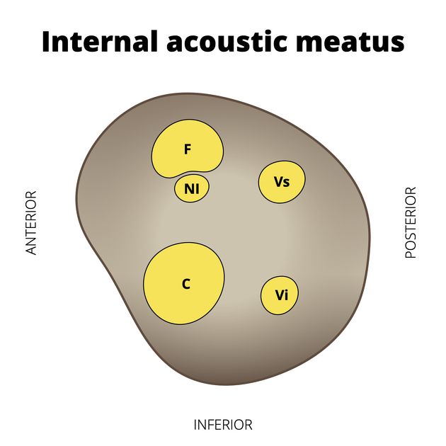
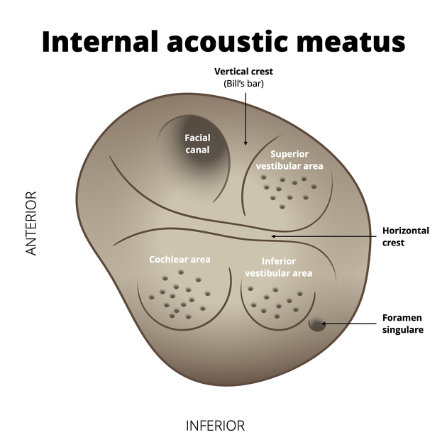
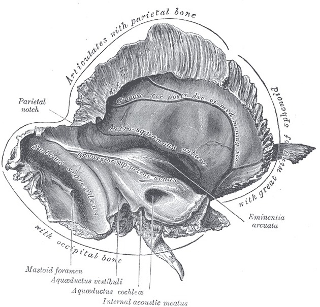
The opening of the IAC, the porus acusticus internus, is located within the cranial cavity, near the posterior surface of the temporal bone. The margins of the opening are smooth and rounded, and the canal is short (1 cm), running laterally to the bone. The canal narrows laterally, and the lateral boundary is the fundus, where the canal splits into three distinct openings, one of which is the facial nerve canal.
Contents
labyrinthine artery (usually a branch of the AICA or basilar artery)
Nerves
There are five nerves that run through the IAC:
nervus intermedius (sensory component of CN VII)
facial motor root (motor component of CN VII)
cochlear nerve (component of CN VIII)
inferior vestibular nerve (component of CN VIII)
superior vestibular nerve (component of CN VIII)
Their position is most constant in the lateral portion of the meatus which is anatomically divided by the falciform crest. This horizontal ridge divides the canal into superior and inferior portions:
superior: facial nerve and superior vestibular nerve (SVN); the facial nerve is anterior to the SVN and is separated from it laterally by Bill's bar, a vertical ridge of bone
inferior: cochlear nerve and inferior vestibular nerve (IVN); the cochlear nerve is situated anteriorly
See mnemonic for the position of the nerves in the IAC.
Ganglion
In addition to the three nerves which enter it, it also contains the vestibular ganglion (ganglion of Scarpa). From here three bundles emerge: superior and inferior division of the vestibular nerve and the nerve from the posterior semicircular canal (see article: vestibulocochlear nerve (CN VIII) for further details).
Variant anatomy
narrow duplicated internal acoustic canal 2
Radiographic features
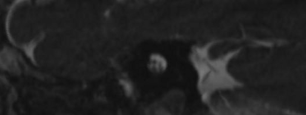
MRI
The anatomy of the IAC is best seen on high-resolution T2-weighted image sequences. Structures that can be seen are facial and vestibulocochlear nerves.
References
- 1. Gray's Anatomy. Churchill Livingstone. (2011) ISBN:0443066841. Read it at Google Books - Find it at Amazon
- 2. Vincenti V, Ormitti F, Ventura E. Partitioned versus duplicated internal auditory canal: when appropriate terminology matters. Otology & neurotology : official publication of the American Otological Society, American Neurotology Society [and] European Academy of Otology and Neurotology. 35 (7): 1140-4. doi:10.1097/MAO.0000000000000458 - Pubmed
Incoming Links
- Sensorineural hearing loss
- Anterior inferior cerebellar artery loop
- Perilymphatic duct
- Vestibular schwannoma
- Internal auditory canal atresia
- Porus acusticus externus
- Möbius syndrome
- Temporal bone (modified Stenvers view)
- Nervus intermedius schwannoma
- Cochlear aqueduct
- Deafness
- Cochlear hypoplasia
- Internal auditory canal nerves (mnemonic)
- Petrous part of temporal bone
- Petrous apex
- Paget disease (bone)
- Cranial foramina
- Anterior inferior cerebellar artery
- Vestibular ganglion
- Porus acusticus internus
- Anterior inferior cerebellar artery loop - type III
- Vestibular schwannoma
- Anterior inferior cerebellar artery loop (Type III)
- Internal auditory canal stenosis
- Skull base hyperpneumatisation
- Intracanalicular vestibular schwannoma
- Trigeminal schwannoma
- Vestibular schwannoma - intracanalicular
- Corpus callosum dysgenesis
- Cerebellopontine angle meningioma
- Epidermoid cyst of the cerebellopontine cistern
- En plaque meningioma with associated CPA / frontal convexity meningiomas
- Meningioma of the cerebellopontine angle
- Meningioma - cerebellopontine angle
- Epidermoid cyst compressing the trigeminal nerve
- Anterior inferior cerebellar artery vascular loop - type 1
- Anterior inferior cerebellar artery vascular loop - type II
- Epidermoid cyst compressing the trigeminal nerve
- Arachnoid cyst: extremely large
- Lipoma-cerebellopontine angle
Related articles: Anatomy: Head and neck
- skeleton of the head and neck
-
cranial vault
- scalp (mnemonic)
- fontanelle
-
sutures
- calvarial
- facial
- frontozygomatic suture
- frontomaxillary suture
- frontolacrimal suture
- frontonasal suture
- temporozygomatic suture
- zygomaticomaxillary suture
- parietotemporal suture (parietomastoid suture)
- occipitotemporal suture (occipitomastoid suture)
- sphenofrontal suture
- sphenozygomatic suture
- spheno-occipital suture (not a true suture)
- lacrimomaxillary suture
- nasomaxillary suture
- internasal suture
- basal/internal
- skull landmarks
- frontal bone
- temporal bone
- parietal bone
- occipital bone
- skull base (foramina)
-
facial bones
- midline single bones
- paired bilateral bones
- cervical spine
- hyoid bone
- laryngeal cartilages
-
cranial vault
- muscles of the head and neck
- muscles of the tongue (mnemonic)
- muscles of mastication
-
facial muscles
- epicranius muscle
- circumorbital and palpebral muscles
- nasal muscles
-
buccolabial muscles
- elevators, retractors and evertors of the upper lip
- levator labii superioris alaeque nasalis muscle
- levator labii superioris muscle
- zygomaticus major muscle
- zygomaticus minor muscle
- levator anguli oris muscle
- malaris muscle
- risorius muscle
- depressors, retractors and evertors of the lower lip
- depressor labii inferioris muscle
- depressor anguli oris muscle
- mentalis muscle
- compound sphincter
-
orbicularis oris muscle
- incisivus labii superioris muscle
- incisivus labii inferioris muscle
-
orbicularis oris muscle
- muscle of mastication
- modiolus
- elevators, retractors and evertors of the upper lip
- muscles of the middle ear
- orbital muscles
- muscles of the soft palate
- pharyngeal muscles
- suprahyoid muscles
- infrahyoid muscles
- intrinsic muscles of the larynx
- muscles of the neck
- platysma muscle
- longus colli muscle
- longus capitis muscle
- scalenus anterior muscle
- scalenus medius muscle
- scalenus posterior muscle
- scalenus pleuralis muscle
- sternocleidomastoid muscle
-
suboccipital muscles
- rectus capitis posterior major muscle
- rectus capitis posterior minor muscle
- obliquus capitis superior muscle
- obliquus capitis inferior muscle
- accessory muscles of the neck
- deep cervical fascia
-
deep spaces of the neck
- anterior cervical space
- buccal space
- carotid space
- danger space
- deep cervical fascia
- infratemporal fossa
- masticator space
- parapharyngeal space
- stylomandibular tunnel
- parotid space
- pharyngeal (superficial) mucosal space
- perivertebral space
- posterior cervical space
- pterygopalatine fossa
- retropharyngeal space
- suprasternal space (of Burns)
- visceral space
- surgical triangles of the neck
- orbit
- ear
- paranasal sinuses
- upper respiratory tract
- viscera of the neck
- blood supply of the head and neck
-
arterial supply
-
common carotid artery
- carotid body
- carotid bifurcation
- subclavian artery
- variants
-
common carotid artery
- venous drainage
-
arterial supply
- innervation of the head and neck
-
cranial nerves
- olfactory nerve (CN I)
- optic nerve (CN II)
- oculomotor nerve (CN III)
- trochlear nerve (CN IV)
-
trigeminal nerve (CN V) (mnemonic)
- trigeminal ganglion
- ophthalmic division
- maxillary division
- mandibular division
- abducens nerve (CN VI)
- facial nerve (CN VII)
-
vestibulocochlear nerve (CN VIII)
- vestibular ganglion (Scarpa's ganglion)
- glossopharyngeal nerve (CN IX)
- vagus nerve (CN X)
- (spinal) accessory nerve (CN XI)
- hypoglossal nerve (CN XII)
- parasympathetic ganglia of the head and neck
- cervical sympathetic ganglia
- greater occipital nerve
- third occipital nerve
-
cervical plexus
- muscular branches
- longus capitis
- longus colli
- scalenes
- geniohyoid
- thyrohyoid
-
ansa cervicalis
- omohyoid (superior and inferior bellies separately)
- sternothyroid
- sternohyoid
- phrenic nerve
- contribution to the accessory nerve (CN XI)
- cutaneous branches
- muscular branches
- brachial plexus
- pharyngeal plexus
-
cranial nerves
- lymphatic drainage of the head and neck
- embryological development of the head and neck


 Unable to process the form. Check for errors and try again.