
Presentation
Work up for hematuria.
Patient Data
Age: 45 years
Gender: Male
From the case:
Renal cell carcinoma


Download
Info
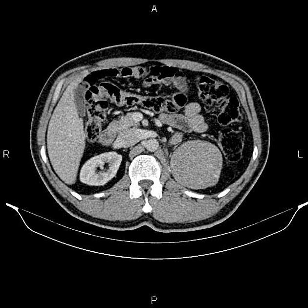
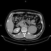
A 75 x 70 mm well-defined low enhancing mass is seen at the upper pole of the left kidney. There is no extension to the adrenal gland or left renal vessels. Infiltration is seen in the pelvicalyceal system.
A few enlarged lymph nodes are noted in the left renal hilum and paraaortic area.
Case Discussion
Left renal mass, pathology proved renal cell carcinoma (RCC) with pelvicalyceal infiltration and regional enlarged lymph nodes.


 Unable to process the form. Check for errors and try again.