
Presentation
Work up for dysphagia and vomiting.
Patient Data
Age: 80 years
Gender: Male
From the case:
Esophagogastric adenocarcinoma


Download
Info
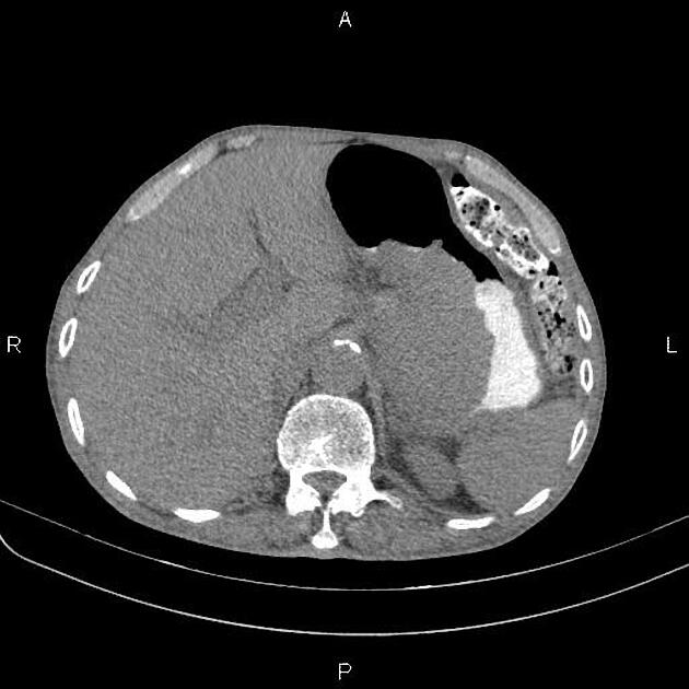
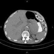
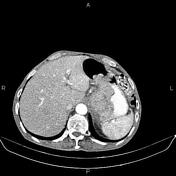
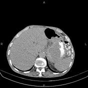
Marked increased wall thickness due to tumoral infiltration is present at the distal esophagus, esophagogastric junction, gastric cardia, and subcardia and proximal of the lesser curvature, accompanied by perigastric fat stranding and several regional enlarged lymph nodes with SAD less than 25 mm. There is no sign of local invasion of the adjacent structures.
Several non enhanced simple cortical cysts are seen at both kidneys.
The prostate gland is enlarged.
A small amount of free fluid is present at the pelvis.
Case Discussion
Esophagogastric mass; pathology proved adenocarcinoma with regional enlarged lymph nodes.


 Unable to process the form. Check for errors and try again.