
Presentation
Blood stained cough.
Patient Data
Age: Adult
Gender: Male

Download
Info
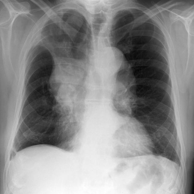
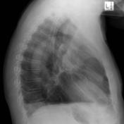
Chest x-ray demonstrates increased density in the right upper medial hemithorax with loss of volume, and shift of the trachea to the right. A mass is present at the right hilum.
Download
Info
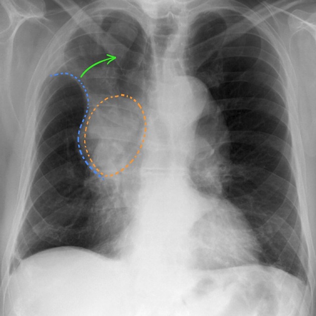
Right hilar mass (orange) obstructing the right upper lobe bronchus results in collapse of the right upper lobe (green arrow). This results in a reverse S shape to the pleural edge.
Case Discussion
This case demonstrates the typical appearances of a reverse S sign of Golden (aka Golden S sign). This patient had a primary bronchogenic carcinoma at the hilum.


 Unable to process the form. Check for errors and try again.