
Presentation
Neonate with abnormal neurology.
From the case:
Type I lissencephaly
Download
Info

Axial slice of non-contrast CT head shows grossly underformed gyri, a smooth cortical surface, band heterotopia and dilated lateral ventricles, more prominent posteriorly (temporal horns) and right more than left. The findings are compatible with lissencephaly type I.
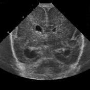
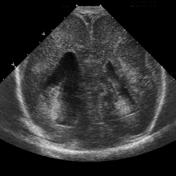
From the case:
Type I lissencephaly

Download
Info

Head ultrasound (HUS) demonstrates the same findings seen on the CT head.
Case Discussion
Typical appearance of lissencephaly type I, with no normal gyration visible, lending a figure 8 appearance to the cortex.


 Unable to process the form. Check for errors and try again.