
- Note: This case has been tagged as "legacy" as it no longer meets image preparation and/or other case publication guidelines.
From the case:
Peritoneal CSF pseudocyst

Download
Info
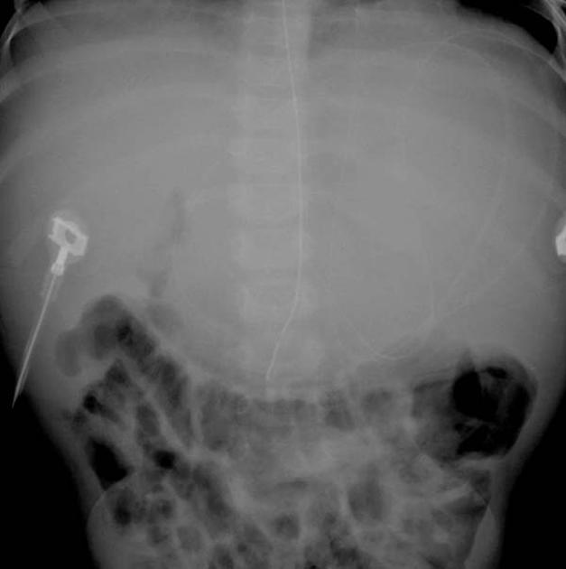
VP shunt catheter in the left upper quadrant, with soft tissue density displacing adjacent bowel.
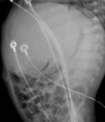
From the case:
Peritoneal CSF pseudocyst
Download
Info
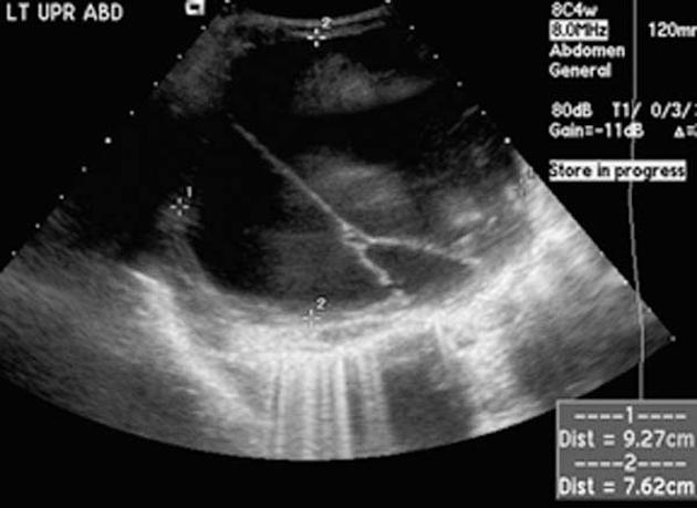
Large multicystic area closely approximated to the shunt catheter tip.
Case Discussion
Features are consistent with a CSF pseudocyst.
This image is from Dr. Paula Brill's excellent pediatric radiology collection.
Dr. Brill is a professor in the department of radiology (pediatric section) at Weill Cornell.
This case was donated to Radiopaedia.org by Radswiki.net


 Unable to process the form. Check for errors and try again.