
Presentation
Renal mass for evaluation
Patient Data
Age: 70 years
Gender: Male
From the case:
Renal cell carcinoma

Download
Info
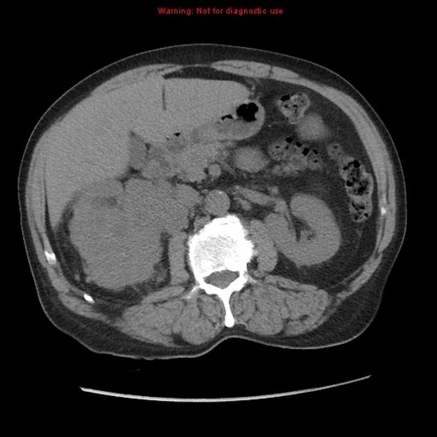
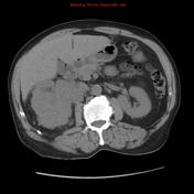
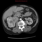
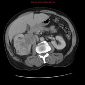
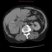
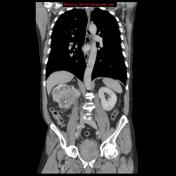
Large right renal mass lesion involving the middle and lower poles with an exophytic component. It is invading the pelvicalyceal system. It shows heterogeneous post contrast enhancement. The upper calyx is compressed with mild dilatation and delayed contrast excretion. Preserved opacification of right renal vein and IVC.
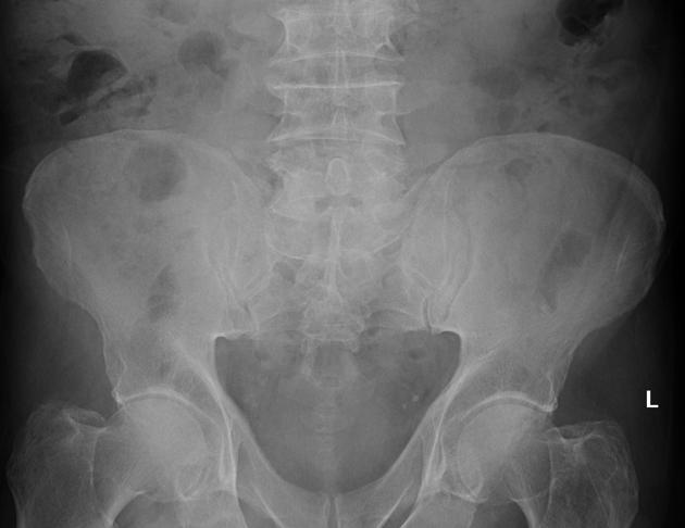
The prostate is markedly enlarged raising the bladder base.
Bilateral direct inguinal hernias, larger on right side.
Case Discussion
Path proven renal cell carcinoma conventional (clear cell) type.


 Unable to process the form. Check for errors and try again.