
Presentation
Acute left flank pain.
Patient Data
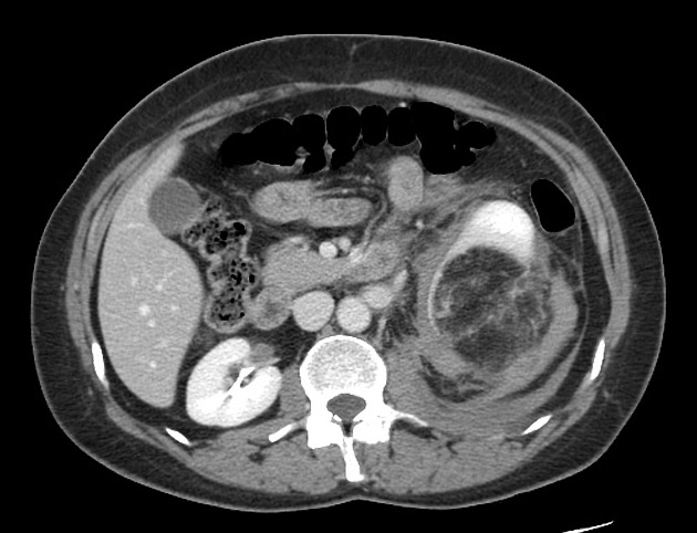
CT of the abdomen demonstrates a large fat density mass arising from the lower pole of the left kidney. It is associated with significant surrounding fluid which extends down towards the pelvis, and up behind the kidney.
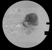
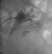
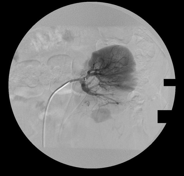
Initial injection into the renal artery demonstrates brisk extravasation of contrast inferiorly, consistent with rapid ongoing bleeding. A microcatheter was navigated into bleeding vessel and glue was used to occlude it (not shown).
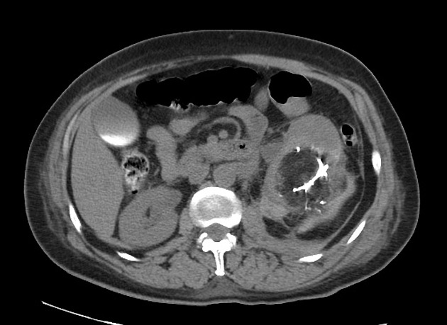
At the inferior pole of the left kidney the a fat-containing soft tissue mass measuring 8.7 x 11.2 x 11.9 is again demonstrated. There is a new curvilinear hyperdense focus within the fat-containing lesion representing sequelae of interval vascular glue embolization. The HU within the soft tissue attenuating peripheral component is approximately 66, probably representing residual hemorrhage.
The proximal ureter is displaced anteromedially resulting in hydronephrosis and proximal hydroureter.
Case Discussion
This case illustrates the most common complication of large angiomyolipomas: spontaneous retroperitoneal hemorrhage.


 Unable to process the form. Check for errors and try again.