
Presentation
Known case of a chronic illness, presented with shortness of breath.
Patient Data
Age: 25 years
Gender: Female
From the case:
Non-specific interstitial pneumonia
Download
Info
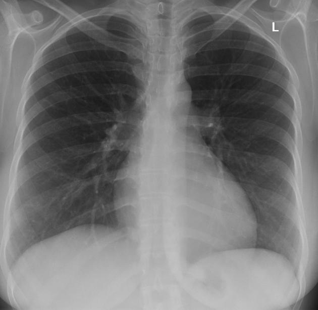
Chest radiograph shows no significant abnormality.
From the case:
Non-specific interstitial pneumonia


Download
Info
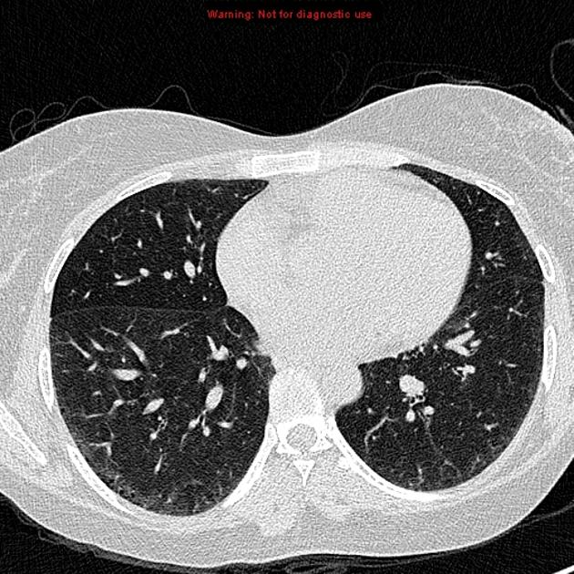
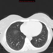
Bilaterally symmetrical fine reticular opacities with relative subpleural sparing, microcystic abnormalities and mild ground glass opacities with lower lobe predominance.
Case Discussion
There is a well known association of NSIP with SLE. In NSIP, fine reticular opacities and microcystic honeycombing represent fibosis while ground glass opacities without traction bronchiectatic changes possibly represent inflammation. It can be normal in early stage of disease and in plain radiographs.


 Unable to process the form. Check for errors and try again.