
Presentation
Acute onset of severe headache.
Patient Data
Age: 50 years
Gender: Male
From the case:
Cerebral arteriovenous malformation
Download
Info

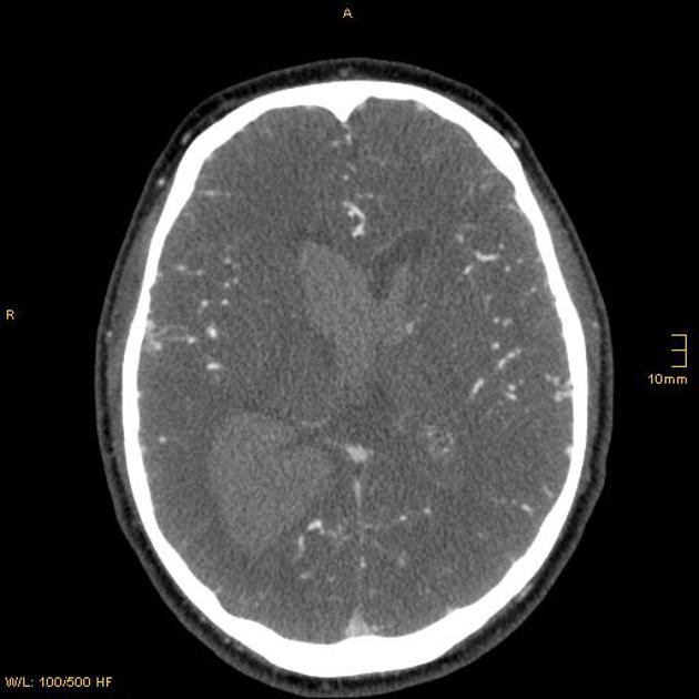
Cranial CT shows intracerebral hemorrhage in the right occipital lobe and extension of parenchymal bleeding into the ventricles.


Download
Info
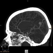
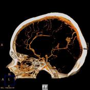
An arteriovenous malformation (AVM) fed by the right posterior cerebral artery and is drained by a single vein into the superior sagittal sinus.
Case Discussion
When an intracranial hemorrhage is seen in an atypical demographic or location an underlying lesion (such as a cerebral vascular malformation or tumor) should be considered and further imaging is often required.


 Unable to process the form. Check for errors and try again.