
From the case:
Aneurysmal bone cyst (ABC)

Download
Info
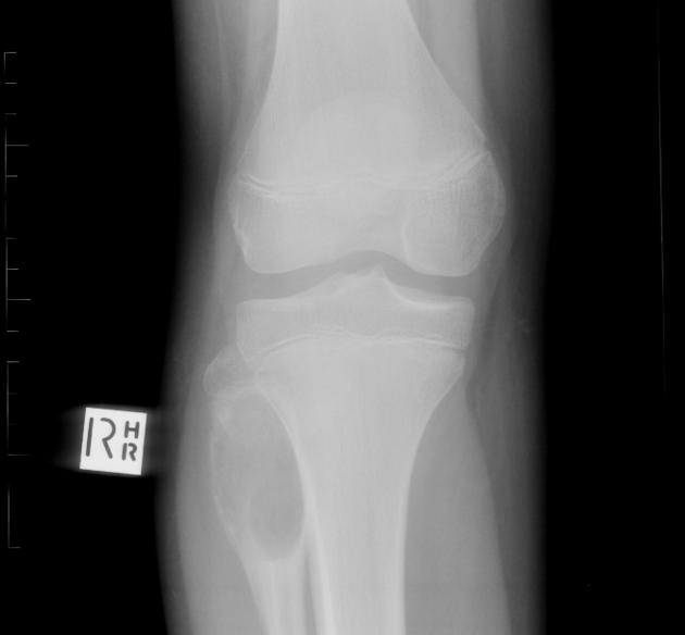
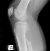
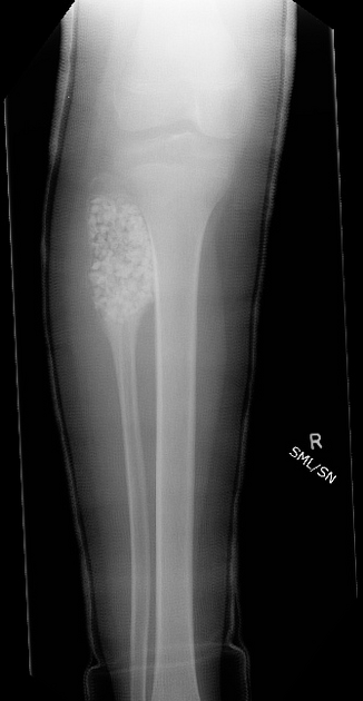
Initial radiographs demonstrate a lucent expansile lesion involving the right neck of fibula.
From the case:
Aneurysmal bone cyst (ABC)
Download
Info
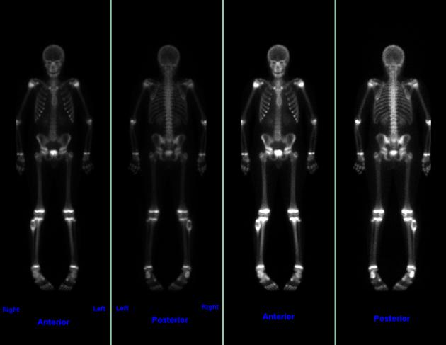
Bone scan demonstrates the "doughnut sign" of aneurysmal bone cyst: peripheral uptake with a photopenic center.
From the case:
Aneurysmal bone cyst (ABC)


Download
Info

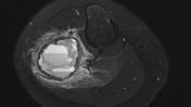
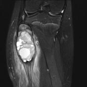
Expansile lesion of the proximal fibular that is T2 hyperintense with numerous fluid-fluid levels. Surrounding soft tissue inflammation.
From the case:
Aneurysmal bone cyst (ABC)

Download
Info
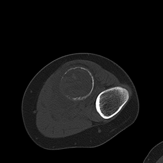
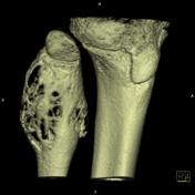
CT demonstrates lytic lesion in the proximal fibular that is expansile with cortical thinning.
Case Discussion
This case demonstrates the multimodality appearance of an aneurysmal bone cyst (ABC).


 Unable to process the form. Check for errors and try again.