
Presentation
This patient had a low grade glioma treated 40 years ago. Now presents with new onset aphasia.
Patient Data


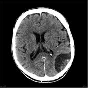
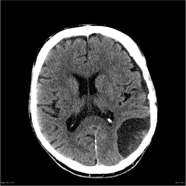
There is a left frontoparietal abnormality with areas of calcification and possible fluid level, measuring approximately 2 cm. This is predominantly surrounded by encephalomalacic changes, with probable minimal parenchymal edema. No abnormal enhancement was detected. There is no convincing evidence of recurrence or residue of a high grade neoplasm at this region, and the appearance is more compatible with chronic post-interventional (surgical +/- radiotherapy) changes.
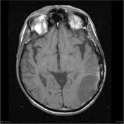
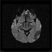
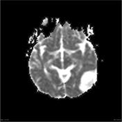
In addition, there is a large left parietotemporal hypoattenuating abnormality, approximately measuring 5 cm, which appears extra-axial. This lesion contains some heterogeneous internal densities with minor enhancement of these foci. There is minimal mass effect with less than 3 mm subfalcine herniation.

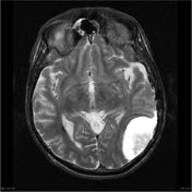

There is evidence of prior left sided craniotomy. Encephalomalacia is seen in the left frontoparietal region with coarse cortically based calcification. In addition there is a 15 mm rounded cystic region containing layering blood products in the region of the left corona radiata.
A large extra axial lesion is seen in the left parietal region measuring 54 mm across the base and 32 mm in height. There is a cystic interface between the lesion and the adjacent cortex and irregular enhancement at the base of lesion. There is moderate mass effect on the adjacent brain.
Moderate white matter disease in the periventricular white matter. Thickening of the mucosa is seen in the paranasal sinuses.
Impression:
- Left parietal extra-axial lesion suspicious for a radiation induced neoplasm, most likely an unusual appearing meningioma, although other radiation induced neoplasms are not excluded.
- Hemorrhagic cystic area in the left corona radiata is noted, most likely relating to prior radiotherapy.
The patient underwent a craniotomy and resection of the extra-axial mass.

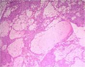
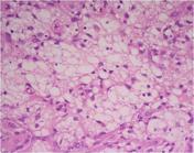
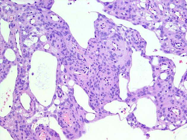
MICROSCOPIC DESCRIPTION: Paraffin sections show a moderately hypercellular meningioma with a mixed syncytial and microcystic architecture. Tumor cells show mild nuclear atypia. Intracytoplasmic cysts are seen, displacing the nucleus peripherally. No mitotic figures or areas of necrosis are identified and there is no evidence of brain invasion. Broad attachment to dura is noted.
DIAGNOSIS: Meningioma - microcystic variant.
Case Discussion
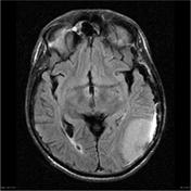
Radiation induced meningiomas are only seen many years after radiotherapy. In this case the microcystic histology contributes to an unusual appearance, with the meningioma being very high in T2 signal. It should be noted that high T2 signal is not pathognomonic for the microcystic variant.


 Unable to process the form. Check for errors and try again.