
Presentation
Left flank pain and hematuria.
Patient Data
Age: 60 years
Gender: Female
From the case:
Renal cell carcinoma


Download
Info

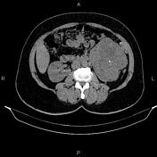
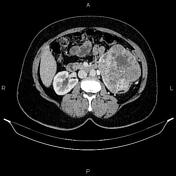
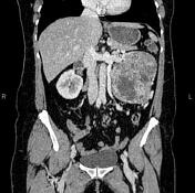
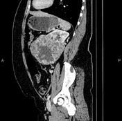
A large exophytic mass is seen in the lower pole of the left kidney, causing a mass effect on the collecting system. A few tiny calcified foci are noted within the mass. After IV contrast media administration, it enhances heterogeneously because of necrotic components.
A few small, simple cortical cysts are seen in the right kidney.
Uterus and ovaries are not seen due to prior resection.
Case Discussion
The patient underwent a left nephrectomy, and histopathology evaluation confirmed renal cell carcinoma (clear cell type).


 Unable to process the form. Check for errors and try again.