
Presentation
Progressive abdominal enlargement
Patient Data
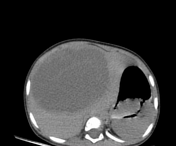

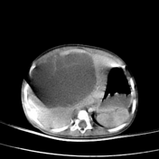

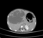
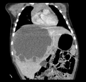
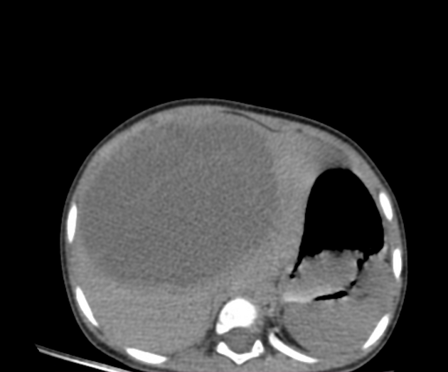
A well-defined large mixed cystic/ solid hepatic lesion is seen occupying most of the liver (Segment IV, V, and VIII respectively). It shows a large septae and a thick wall giving a Swiss chees appearance. After IV contrast, it shows heterogeneous enhancement of its solid component. It measures 8.4 x 10.2 x 11 cm (AP x Tr and CC respectively).
Gross appearance: specimen received was 13 x 9 x 7 cm with a smooth outer surface. C/S was multilocular containing serous and gelatinous material with a thin wall and hepatic tissue. The surgical margin was about 3 mm processed in block (S), and the lesion was processed in block C.
Microscopic examination: Revealed a lesion formed of loose myxoid connective tissue with dilated vessels and proliferating epithelial bile ducts arranged in lobulated islands. Some of these ducts are arranged in aductal plate malformation pattern. Other foci show collagenous concentric whorls around some ducts. There are wide cystic structures filled with light pink fluid. Islends of hepatocytes are present in the wall of cyst. No detected atypical cells.
Final diagnosis: Picture consistent with mesenchymal hamartoma.
Courtesy to Prof. Dr. Khadiga Ali professor of pathology, Mansoura faculty of medicine.
Case Discussion
The patient underwent complete tumor resection and was pathologically proven to be a hepatic mesenchymal hamartoma.
Hepatic mesenchymal hamartoma is a rare hepatic tumor (8%) of the pediatric age group below the age of 2 1.
It has several differential diagnoses with other pediatric liver masses. the most common appearance on CT, is a mixed solid/ cystic hepatic mass with enhancing septae.


 Unable to process the form. Check for errors and try again.