
Presentation
Slowly growing non-painful left lateral neck swelling.
Patient Data


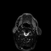
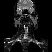
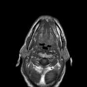
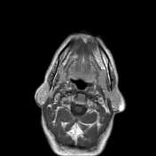
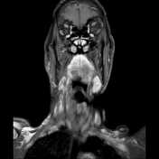
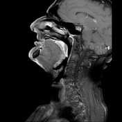
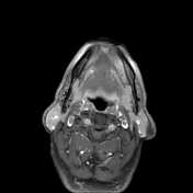
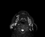
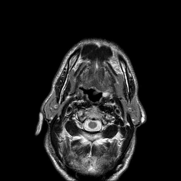
A well-encapsulated soft tissue mass is seen at the left lateral neck region superficial to the parotid tail, measuring 2.9 x 1.7 x 3 cm (Ap X Tr and CC respectively). It elicits high signals on both T1 and T2 images with suppression on fat suppression sequences. It shows a thin peripheral enhancement. No definite deeper or intramuscular extension.
Left cerebellar developmental venous anomaly is noted (incidental finding).
Case Discussion
The MRI findings are highly suggestive of left neck lipoma.
MRI is an excellent technique for assessing soft tissue lipomas. Classically, they display high signals on both T1 and T2 images with saturation on fat-saturated sequences (STIR, SPAIR and T1 fat suppression). It is also valuable to exclude low-grade liposarcoma which has thick enhancing septae and heterogeneous signal.


 Unable to process the form. Check for errors and try again.