
Presentation
Prolonged cough with elevated inflammatory markers.
Patient Data
Age: 50 years
Gender: Male
Download
Info
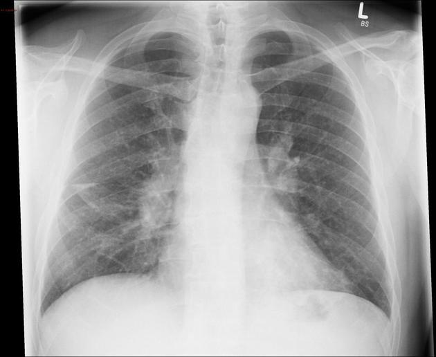
Consolidation on the right and hilar structure raised suspicion and lead to CT scan. Also note the left lower lobe pneumonia.


Download
Info
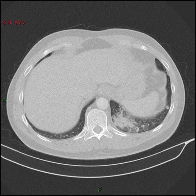
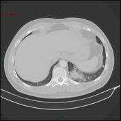
Pneumonic infiltration in the left lower lobe and right middle lobe. Also nodular consolidation in the left lower lobe and micronodular densities throughout. Mediastinal and hilar lymphadenopathy.


 Unable to process the form. Check for errors and try again.