
Presentation
3 days of purpuric rash, hematemesis and diarrhea.
Patient Data
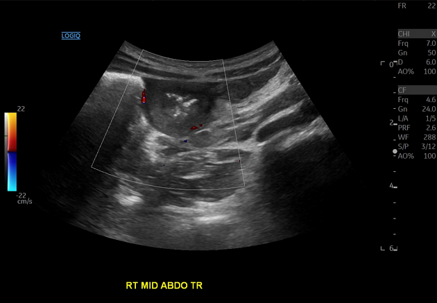
FINDINGS: The visualized proximal duodenum displays circumferential mural thickening, single wall thickness 7 mm. Outer wall to outer wall diameter is 29 mm.
Remainder of the imaged bowel is normal in appearance.
Small volume of free fluid, as seen on previous ultrasound.
IMPRESSION: 1. Circumferential mural thickening of the proximal duodenum. This likely represents intramural hemorrhage secondary to IgA vasculitis (Henoch-Schonlein purpura), particularly given pre-existing clinical diagnosis of this condition. Differential includes an inflammatory or infectious process, considered less likely.
2. No evidence of intussusception.
Transferred to pediatric rheumatology service. Diagnosis of IgA vasculitis was confirmed based on clinical features, abdominal US, and skin biopsy findings. Treated with steroids.
Case Discussion
IgA vasculitis (Henoch-Schonlein purpura) gives rise to gastrointestinal symptoms in one half of affected children, due to submucosal hemorrhage and edema. Most common sites are duodenum, stomach and colon. Intussusception is the commonest GI complication.


 Unable to process the form. Check for errors and try again.