
Presentation
Incidental finding on ultrasound.
Patient Data
Age: 70 years
Gender: Male
From the case:
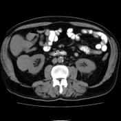
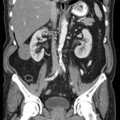
Hypovascular renal cell carcinoma


Download
Info

5 cm right renal mass lesion isodense to renal parenchyma. Weak contrast enhancement from 40 to 50 HU on the average.
Case Discussion
Histology: Papillary renal cell carcinoma type I (pT2).


 Unable to process the form. Check for errors and try again.