
Presentation
Mild respiratory distress.
Patient Data
Age: 1 day
Gender: Male
From the case:
Congenital pulmonary airway malformation (CPAM)

Download
Info

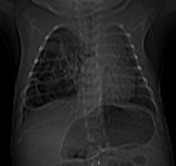
The scout view shows cystic areas in the right hemithorax with contralateral mediastinal shift. The right hemidiaphragm is intact.
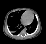
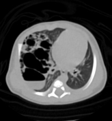
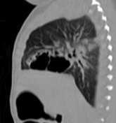
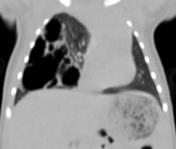
The CT scan (lung window) shows multiple cysts of various sizes (up to 3 cm) involving mainly the right upper lobe, the largest shows an air-fluid level with leftward mediastinal shift. No surrounding lung consolidation. The right hemidiaphragm is intact. The left lung is clear.
Case Discussion
CT features of multiple lung cysts of various sizes (up to cm) in a newborn most consistent with congenital pulmonary airway malformation (CPAM) type 1 according to Stocker classification.


 Unable to process the form. Check for errors and try again.