
Presentation
Workup for chest pain, hemoptysis, and cough.
Patient Data
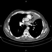

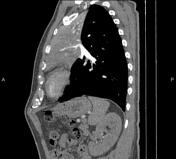


There is low-attenuation collapse consolidation containing enhanced branching vessels in the left upper lobe and lingula segments that resemble CT angiogram sign.
The mass infiltrates the left lung hilum, and several enlarged pre-vascular and aortopulmonary window lymph nodes are noted with SAD less than 15 mm.
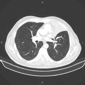
In addition, several small subpleural nodules are also seen in both lungs less than 8 mm.
Case Discussion
Pathology-proven adenocarcinoma in an elderly patient with chest pain, hemoptysis, and cough.
The CT angiogram sign refers to normal vessels on contrast-enhanced CT as they traverse an airless low-attenuation portion of consolidated lung.
Although lung adenocarcinoma is the primary diagnosis, other benign and malignant lesions such as infectious and post-obstructive pneumonia, pulmonary edema, pulmonary lymphoma, and metastasis from gastrointestinal carcinoma also can cause similar appearances on post-contrast CT images.


 Unable to process the form. Check for errors and try again.