
Presentation
Rectal bleeding and abdominal pain. Endoscopy & biopsy-proven case of "Crohn's disease" since 7 years ago.
Patient Data
Age: 35 years
Gender: Female
From the case:
Crohn disease


Download
Info
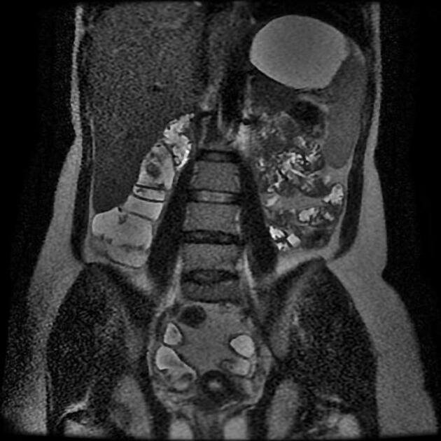
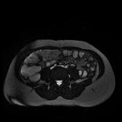
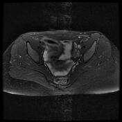
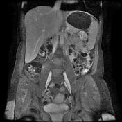
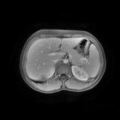
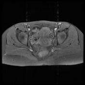
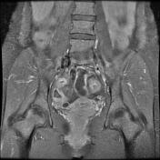
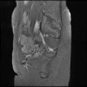
mural thickening and contrast enhancement of the mucosa of the ileal loops and terminal ileum
prominent peri enteric vasculature of the mesentery
vascular dilatation and tortuosity of the vasa recta, giving the comb sign
Case Discussion
Crohn's disease is an idiopathic inflammatory bowel disease (IBD), characterized by widespread discontinuous gastrointestinal tract inflammation. The terminal ileum and proximal colon are most often affected. The comb sign refers to the hypervascular appearance of the mesentery in active Crohn's disease.


 Unable to process the form. Check for errors and try again.