
Presentation
Headache and altered level of consciousness.
Patient Data
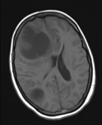

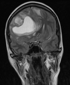
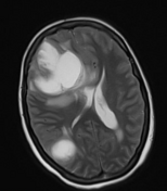
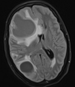
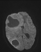
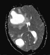
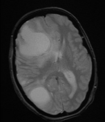

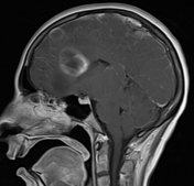
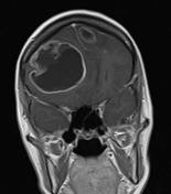

There are innumerable variable-sized intra-axial brain lesions seen in both cerebral, cerebellar hemispheres and left pons with their epicenter close to the grey-white matter junction. They show hypointense signal on T1, hyperintense signal on T2 with hyperintense perilesional edema of variable amounts and intense post-contrast ring enhancement with facilitated diffusion in the center of the lesions.
The largest lesion appears lobulated and is seen in the right frontal lobe with significant perilesional vasogenic edema causing mass effect upon the corpus callosum and the frontal sulci, which appear effaced, resulting in subfalcine herniation. Pending right-sided uncal herniation is noted.
Case Discussion
This patient has an established history of metastatic breast cancer. The features are highly suggestive of brain metastases.


 Unable to process the form. Check for errors and try again.