
Presentation
Testicular seminoma treated by left-sided orchofuniculectomy and three cycles of chemotherapy.
Patient Data


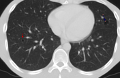
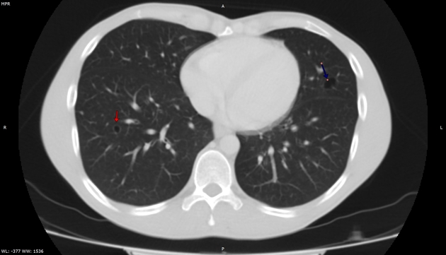
In the VIII segment of the right lung and in the VI segment of left lung there are cystic lesions measuring 0.7 cm and 1.3 cm respectively.

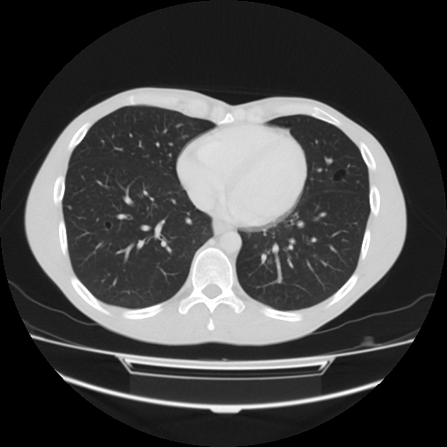
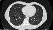
The cystic lesion in the right lung has increased almost 3 times from 0.7 cm to 2.0 cm. There are new areas of focal wall thickening.
The air cyst in the left lung is stable.
There are no other distant metastases.
The patient refused further chemotherapy.

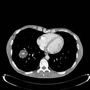

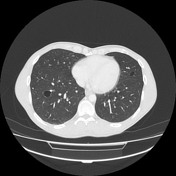
There is now a 4 cm mass containing small cavities at the site of the right segment VIII lesion.
The left lung cyst is stable.
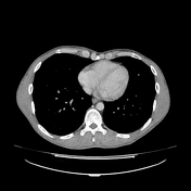
There are new widespread lung, kidney, and liver lesions and lesions in the pancreas and right adrenal gland.

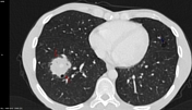
Single slices from the 3 CT scans in chronological order show:
Cyst in the right lung (red arrow),
Cyst in the left lung (blue arrow).
After 5 months, the right lung lesion has increased 3 times.
After 3 years there is a mass at the location of right lung lesion measuring 4 cm. The left lung cyst remains stable.
Case Discussion
Cystic pulmonary metastases are very rare. The main differential diagnosis for it are blebs and bullae. Increased size and wall thickening are suspicious of tumor. At 3 years the cystic lesion has been subsumed by a mass, compatible with metastasis. Metastatic disease is likely although not confirmed by biopsy. The new metastatic lesions in the lungs, kidneys, liver, pancreas, and right adrenal gland add support to this impression.


 Unable to process the form. Check for errors and try again.