
Presentation
Painless, palpable masses in both thighs and around the knee joints.
Patient Data
Note: This case has been tagged as "legacy" as it no longer meets image preparation and/or other case publication guidelines.
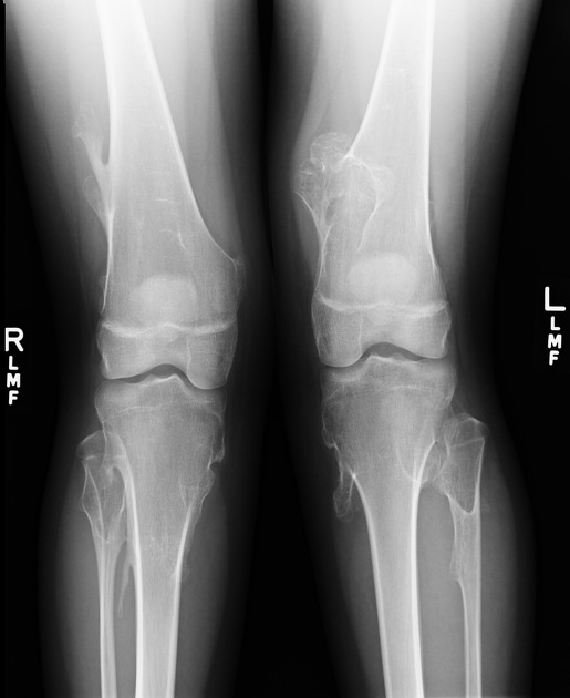
Multiple exostoses of the proximal and distal femora, as well as the proximal tibiae and fibulae. The plain radiograph of both knees demonstrates the numerous exostoses bilaterally.

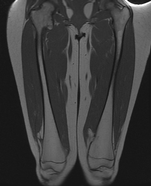
After a plain radiograph, MRI was performed.
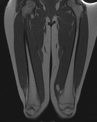
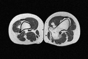
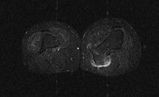
Multiple exostoses of the proximal and distal femora, as well as the proximal tibiae and fibulae. The largest left sided exostosis is seen posteromedially in the distal femoral metaphysis in the coronal and axial T1W MRI images. The hamstring and pes anserine muscles and popliteal neurovascular structures are displaced by this large exostosis. The cartilaginous cap measures 4mm thick, as seen in the axial T2W MRI image.
Case Discussion
Multiple hereditary exostoses (MHE) is an autosomal dominant disorder manifested by multiple osteochondromas, which are benign tumors projecting from the surface of bones. The incidence of MHE is 1 in 50,000 individuals. The exostoses are either sessile or pedunculated, and are located at or close to the metaphyses of the bones involved. Although osteochondromas are benign lesions, they sometimes cause skeletal deformities which may in turn cause clinical symptoms. Short stature is present in 40% of patients. The painless masses can become painful when they impinge upon tendons, muscles, or nerves. Other symptoms include bursitis, fractures, premature osteoarthrosis, weakness or numbness due to impingement of an adjacent nerve, blood vessel aneurysms, and loss of range of motion.
The most serious complication of MHE is the potential for an exostosis to undergo malignant transformation into chondrosarcoma. In skeletally mature patients, the cartilaginous cap should be measured. If the cap is >1 cm thick, there is an increased risk for malignancy. T2-weighted MRI images are the most useful in evaluating the cartilaginous cap.


 Unable to process the form. Check for errors and try again.