
Presentation
Underwent ventriculoperitoneal shunt insertion for aqueductal stenosis, now presenting with enlarging abdomen.
Patient Data
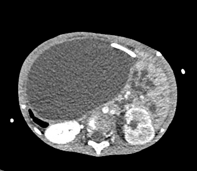
There is a septated irregularly shaped cystic mass occupying the abdominopelvic cavity, measuring 16.9 x 10.8 x 7.6 cm or 725 cc. The ventriculoperitoneal shunt courses within. It displaces the surrounding viscera, bowels, and vessels.
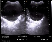
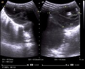

Ultrasound shows a 743 cc septated intraperitoneal fluid collection. The ventriculoperitoneal shunt was seen coursing within the collection.
Case Discussion
Peritoneal pseudocyst formation is a rare complication of ventriculoperitoneal shunt, with reported incidence of up to 4.5%. These cystic masses are lined by fibrous tissue, without epithelial lining. Visualization of the shunt tip within the cystic mass is helpful for establishing the diagnosis. Other imaging features include the presence of septations, irregular shape due to indentations from surrounding structures, and homogeneous low density on CT or anechoic fluid on ultrasound.


 Unable to process the form. Check for errors and try again.