
Presentation
Blindness, leukocoria on examination, excessive tears.
Patient Data
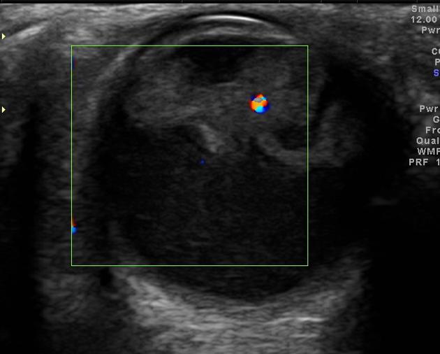
Low level echoes in the vitreous are seen with irregular echogenic soft tissue in retrolental area, with a central arterial flow.


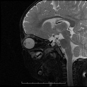

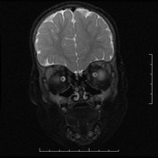
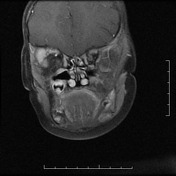
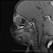
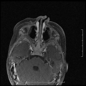

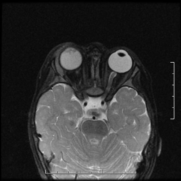
Mildly hyperintense signal vitreous is seen, with no appreciable post-contrast enhancement. A central hyaloid artery is seen extending from lens to retina, giving a characteristic 'wine-glass' appearance. Right lacrimal gland enlargement was also noted.

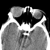
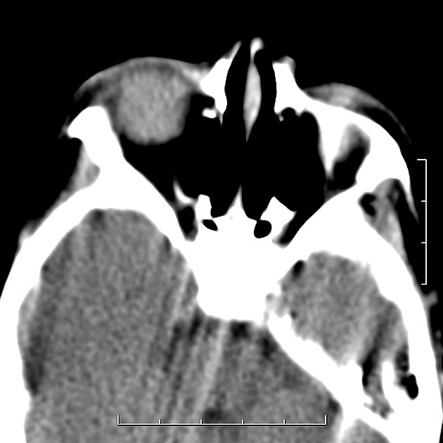
Hyperdense vitreous, with no evidence of any calcification
Case Discussion
Persistent hyperplastic primary vitreous (PHPV) is a rare developmental disorder of childhood. It is one of the differentials of leukocoria, which should be ruled out. Characteristic presence of persistent hyaloid artery, and absence of contrast enhancement or calcification, differentiates it from other conditions like retinoblastoma or Coat's disease.
Lacrimal gland enlargement and PHPV does not seem to have a direct association, however does explain excessive tears in this patient.


 Unable to process the form. Check for errors and try again.