
Presentation
Left upper limb weakness.
Patient Data


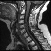
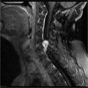

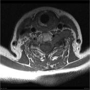
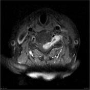
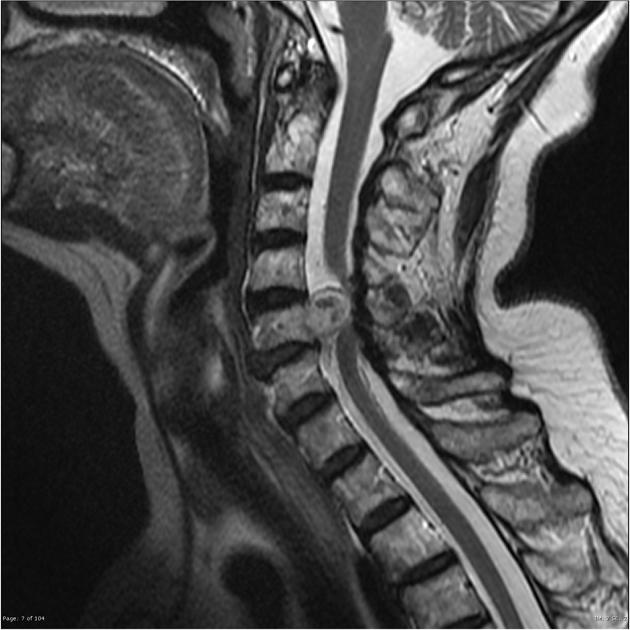
A vividly enhancing dumbbell shaped mass extends through the C5/6 neural exit foramen along the C6 nerve root on the left. It is hyperintense on T2 weighted imaging and isointense to cord on T1. Spinal cord compression with displacement to the right by the mass.
The bony margins of the foramen are remodeled and expanded.
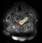
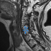
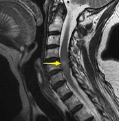
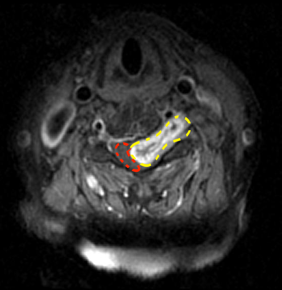
Dumbbell shaped left sided mass (yellow dotted line) passes out of the neural exit foramen which it markedly expands (blue arrows). The cord is displaced towards the right (red dotted line) and has faint increased T2 signal within its substance (yellow arrow).
Histology
MICROSCOPIC DESCRIPTION: Paraffin sections show a moderately hypercellular tumor composed of a mixture of spindle cells and cells with small round hyperchromatic nuclei and pale cytoplasm. Both cell types are arranged in diffuse sheets. Collections of hemosiderin-filled macrophages are identified. Foci of dystrophic calcification are noted. No mitotic figures are identified and there is no necrosis. Immunohistochemistry shows strong staining in tumor cells for S-100 protein and nestin. No staining for epithelial membrane antigen (EMA), progesterone receptor or E-cadherin is seen in tumor cells.
FINAL DIAGNOSIS: Spinal Neurilemmoma (Schwannoma).
Case Discussion
This case illustrates fairly typical appearances of spinal schwannoma extending along the C6 nerve root.


 Unable to process the form. Check for errors and try again.