
Presentation
Abdominal pain and weight loss.
Patient Data
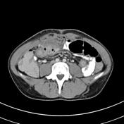


There is a long segment of distal small bowel thickening along with engorgement of the adjacent mesenteric vessels.
Around the level of umbilicus, midline anteriorly and just deep to the recti muscles, there is a peripherally enhancing collection containing gas locules, surrounded by inflammatory stranding.
Colon is unremarkable. Upper abdominal solid viscera look normal. No retroperitoneal lymph node enlargement. Incidental GB stones. No features of acute cholecystitis.
Interpretation: Features are highly suspecious of inflammatory bowel disease (Crohn's) complicated by intra-abdominal abscess.
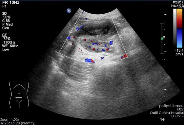
Oval hypoechoic abnormality anteriorly in the abdomen, surrounded by an area of hyperemia. Representing abscess.
GB stones again noted.
Case Discussion
Crohn's disease complicated by intraabdominal abscess.


 Unable to process the form. Check for errors and try again.