
Presentation
Acute onset lower back pain radiating into his right leg. On examination, the patient has scoliosis.
Patient Data
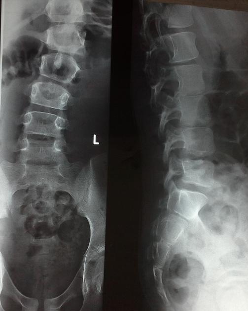
AP and lateral view of the spinal column reveals scoliosis with a sharp angular bend associated with posterior scalloping of the vertebral bodies.
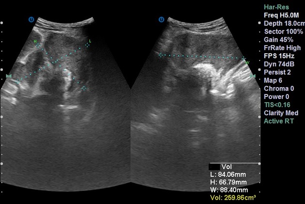
Transverse US image shows a retroperitoneal lobulate border heterogenous echogenicity mass with average volume of 260 cc.
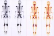
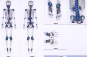
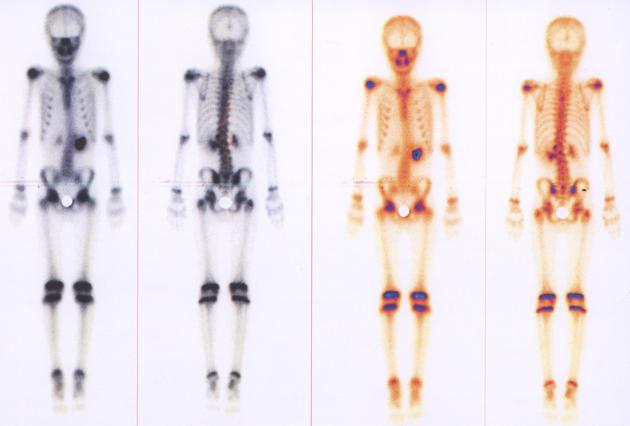
On the dynamic phase,normal perfusion and blood pool activity was noted at the region of lumbar and thoracic spine. On the static phase,severe lumbar scoliosis with focal heterogeneous increased uptake of L1 to L3 as well as T4 were detected. Other parts of skeletal system revealed normal activity.There is retention of activity in the region of left kidney.


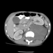
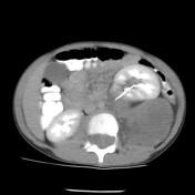
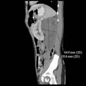
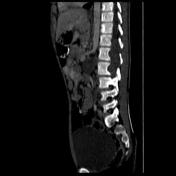
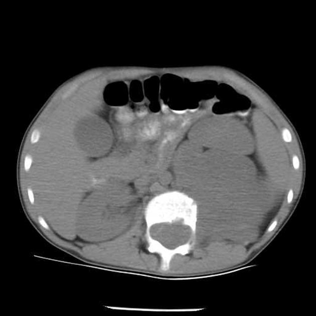
Fusiform longitudinal soft tissue density mass is present against T12 - L2 vertebrae associated with extension in to the spinal canal which causes canal widening and posterior scalloping of mentioned vertebrae. Scoliosis with convexity to the left side is noted. There are also infiltration of left iliopsoas muscle and anterior displacement of left kidney. In addition there are bilateral widening of neuoroforamina in T12-L2 vertebrae.
Case Discussion
Findings are consistent with neurofibromatosis and sudden onset of patient's symptoms is suggestive of neurofibrosarcoma. Patient went on to have a biopsy, but histopathology confirmed ganglioneuroma.


 Unable to process the form. Check for errors and try again.