
Presentation
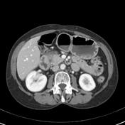
This patient had a history of alcohol-induced pancreatitis. Endoscopic ultrasound was concerning for a pancreatic head mass. Histology of samples taken was inconclusive
Patient Data


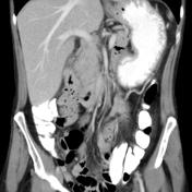
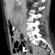
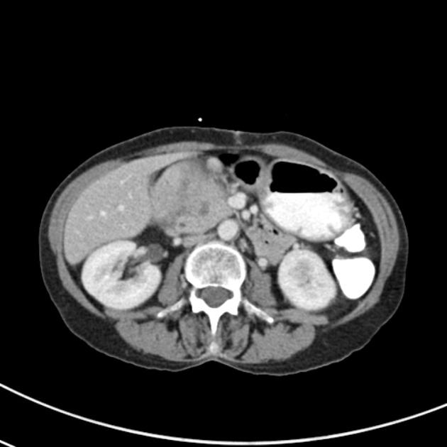
The pancreatic head is expanded with associated peripancreatic inflammatory change, in particular involving the pancreaticoduodenal groove. Within the pancreatic head, there is a 1.1 cm apparently cystic lesion with a possible internal septum. There is no biliary ductal dilatation. The pancreatic duct is dilated (4mm) and somewhat irregular along its course to the level of the pancreatic head beyond which it is no longer visible. The gallbladder is non-distended. Fat planes surrounding the major upper abdominal vascular structures are unremarkable. The portal vein and SMV are normal in caliber without focal narrowing. There is no upper abdominal lymphadenopathy. No focal liver lesions.

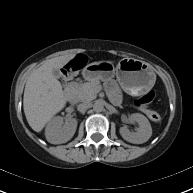
Follow-up examination 3 months later to monitor treatment response, and to exclude an underlying mass.
There has been interval reduction in size of the pancreatic head with reduction in the degree of peripancreatic inflammatory change. The maximal diameter of the pancreatic head currently measures 3.5 x 3.0 cm compared with 4.7 x 4.0 cm previously.
There is hypodense material in the pancreaticoduodenal groove and extending into the pancreatic head. There is some infiltration of the fat plane along the anterior surface of the pancreatic head which has reduced in the interim since the prior study.
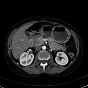
The previously noted discrete cystic pancreatic head lesion is no longer apparent however the pancreatic parenchyma in the head appears heterogeneous with focal areas of low attenuation. The pancreatic duct remains mildly dilated (4 mm). There is no biliary ductal dilatation. The fat planes are preserved surrounding the celiac, SMA and hepatic arteries. The portal vein splenic vein and SMV the normal. No concerning upper abdominal lymphadenopathy. No focal liver lesions.
Case Discussion
This uncommon form of chronic pancreatitis can mimic pancreatic carcinoma. The potential space between the pancreatic head, the common bile duct and the duodenum is referred to as the pancreaticoduodenal groove.
In this case, the overall impression was that of improving paraduodenal pancreatitis. On follow-up CT there was no evidence of a mass, but the pancreatic head remained heterogenous in appearance.


 Unable to process the form. Check for errors and try again.