
Presentation
Large mass medially in the proximal right arm. Recent rapid change in size with clinical deterioration of the patient.
Patient Data
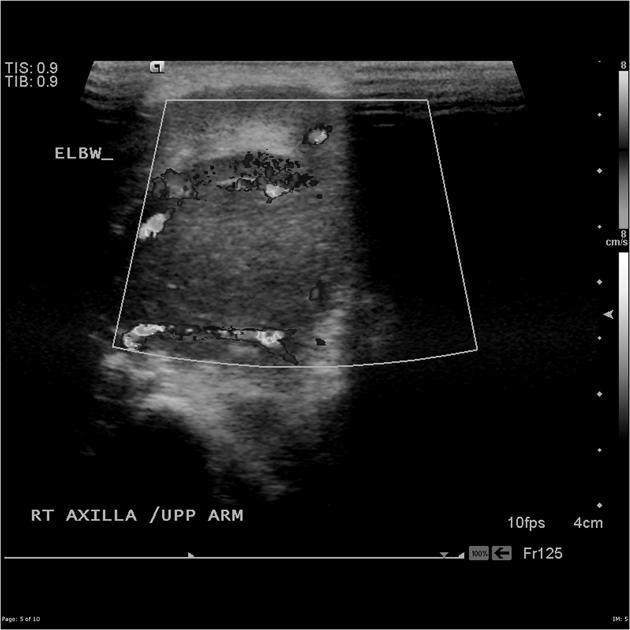
Medially in the proximal upper arm is a large heterogeneous hypoechoic tumor with internal vascularity.
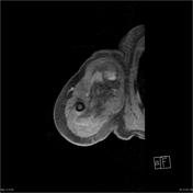

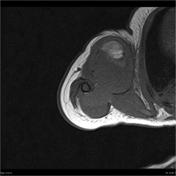
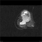
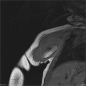
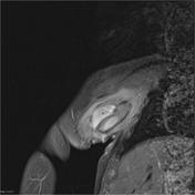

Medially in the right upper arm is a huge high-T2, low-T1 tumor avidly enhancing tumor with a large high-T1 region that has increased signal post-contrast.

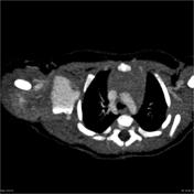
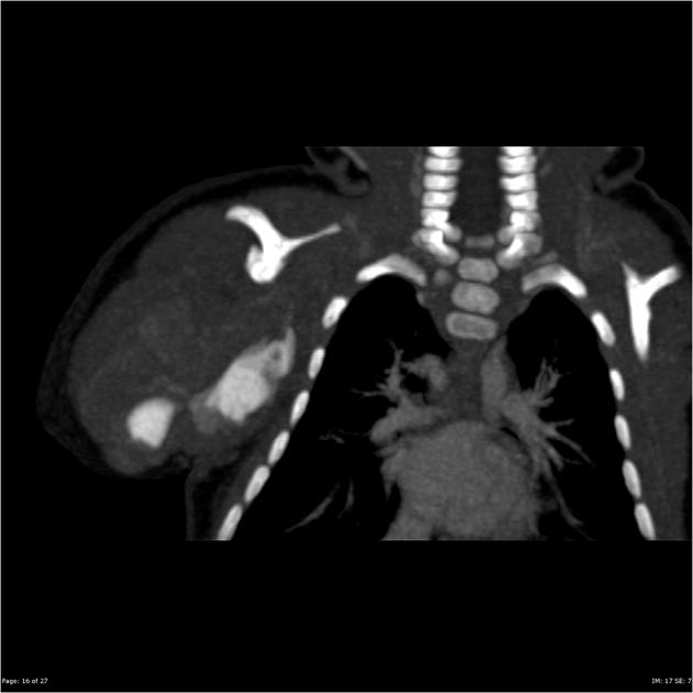
Within the central tumor, there is active extravasation of contrast with pooling of contrast and layering on the CT.
Case Discussion
The rapid change in size and clinical deterioration is attributable to hemorrhage into the tumor which has eroded into an axillary vessel.
Embolization controlled the bleeding. Biopsy confirmed the diagnosis of infantile fibrosarcoma.


 Unable to process the form. Check for errors and try again.