
Presentation
Found intoxicated and submerged in a canal.
Patient Data
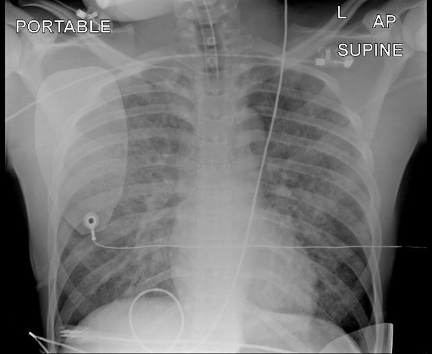
The patient is intubated. There are bilateral airspace opacities with peripheral sparing. Heart and mediastinal contours appear normal.
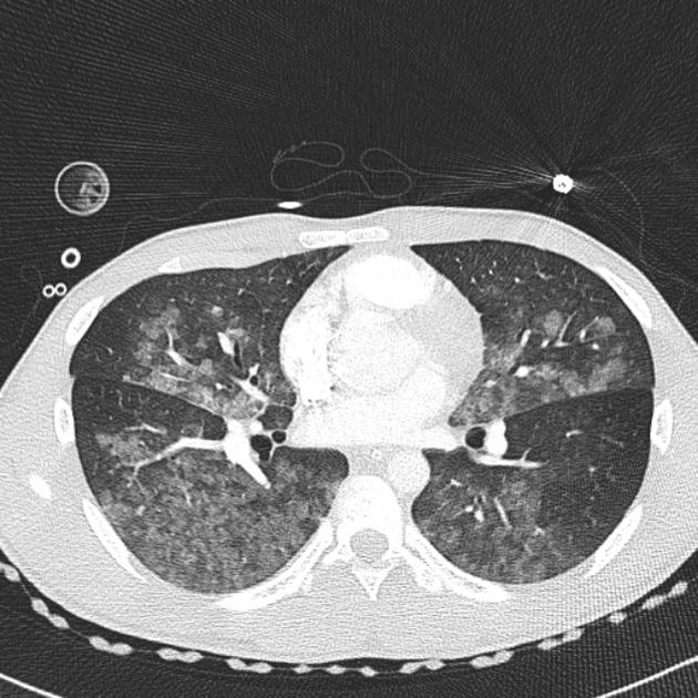
Patchy airspace opacification extending to pleural surfaces posteriorly and at the fissures with sparing of anterior zones. No pleural effusion.

There has been progression of airspace opacities The patient remains intubated and on ventilatory support.
Case Discussion
This type of non-cardiogenic pulmonary edema is considered a result of inhalation of either fresh- or seawater. The radiographic signs are non-specific and resemble those of any other cause of pulmonary edema.
This patient was weaned off ventilatory support and left ICU after 5 days. He did not suffer hypoxic-ischemic brain injury.
This case also shows an inherited radiological misnomer that is likely to continue into perpetuity: the image is labeled "portable" which is patently incorrect. The machine that generated the image is mobile but not portable. You cannot pick it up but you can move it around the hospital.


 Unable to process the form. Check for errors and try again.