
Presentation
Jaundice
Patient Data
Age: 30 years
Gender: Female
From the case:
Choledocholithiasis on CT and MRI


Download
Info

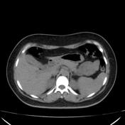
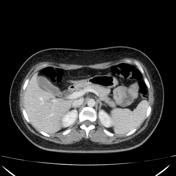
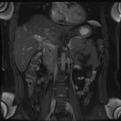
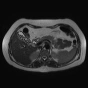
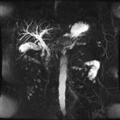
The gallbladder, contains hyperdense bile, with dilatation of the extrahepatic bile duct. The pancreas is edematous.
From the case:
Choledocholithiasis on CT and MRI


Download
Info
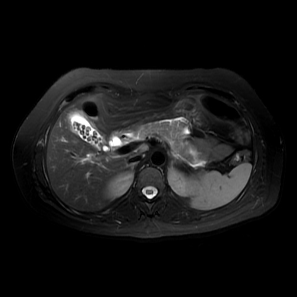
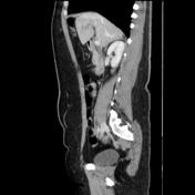
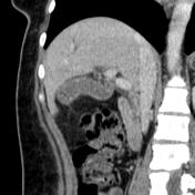
There are multiple calculi within the gallbladder and common bile duct. The pancreas is enlarged and with mild peripancreatic fluid.
Case Discussion
This case shows choledocholithiasis causing mild acute pancreatitis


 Unable to process the form. Check for errors and try again.