
Presentation
This patient has vague abdominal discomfort and was investigated with elective CT abdomen
Patient Data


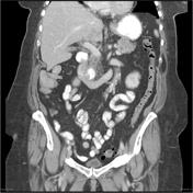
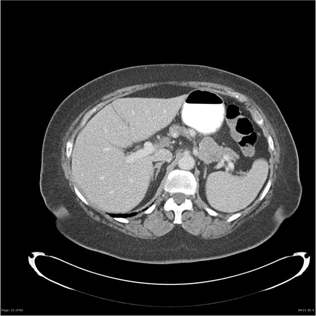
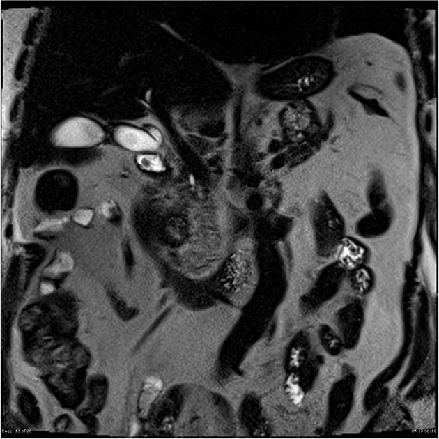
Mildly heterogeneous 3.8 x 3.2 cm pancreatic tail mass centered between the stomach and left adrenal gland. The main pancreatic duct is not dilated. Incidental duodenal diverticulum also shown


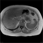
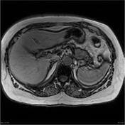
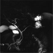

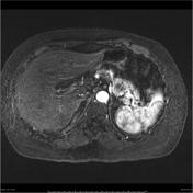
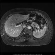
There is a solitary 3.3 cm x 3.6 cm x 2.9 cm (craniocaudal) slightly lobulated T2 hyperintense mass at the superior aspect of the pancreatic tail, abutting the posteroinferior stomach wall. The MRCP sequence reveals the mass to consist of tiny cystic spaces throughout. The main duct is not dilated and does not appear to communicate with the mass. The solid elements of the mass enhance to the same degree as remaining pancreatic parenchyma.
A 2 cm duodenal diverticulum is also present. The biliary tree is normal in appearance. Some dependent filling defects are shown in the gallbladder neck in keeping with stones. There is also a waisted appearance of the gallbladder and focal thickening of the gallbladder fundus containing cystic spaces, diagnostic of focal adenomyomatosis. Borderline normal 9.5 mm short axis lymph node at the porta. No free fluid. Spleen, adrenal glands, kidneys (cysts) appear unremarkable. Flat lipoma deep to left external oblique.
Conclusion:
The pancreatic tail mass is compatible with a serous cystadenoma. Incidental gallstones, duodenal diverticulum and gallbladder adenomyomatosis.
Case Discussion
A female in her 70s is the typical patient for this benign lesion. In this case, the serous cystic adenoma is of the microcystic variant. Even on CT, some small cysts can be appreciated. The tumor can look like a branch-duct IPMN, however unlike IPMNs it has no communication with the ductal system.
Serous cystic tumor is the only pancreatic cystic tumor that is not premalignant.
Incidental adenomyomatosis of gallbladder is shown.


 Unable to process the form. Check for errors and try again.